39 labeled diagram of compound microscope
Looking at the Structure of Cells in the Microscope A light microscope. (A) Diagram showing the light path in a compound microscope. Light is focused on the specimen by lenses in the condensor. ... (Figure 9-15); labeled with electron-dense particles such as colloidal gold spheres, they are used for similar purposes in the electron microscope (discussed below). Ternary Phase Diagram - an overview | ScienceDirect Topics A point on the diagram represents a composition that is specified in terms of mole fraction or weight fraction. The point, (0.3, 0.4, 0.3) is at the center of the small triangle in the diagram and is located by following the red diagonal 60° line at red 0.3 and the horizontal line at blue 0.4 or any combination of two of the coordinates (A, B, C).
Parts of Stereo Microscope (Dissecting microscope) - labeled diagram ... If you would like to learn optical components of a compound microscope, please visit Compound Microscope Parts - Labeled Diagram and their Functions, and this article. How to use a stereo (dissecting) microscope. Follow these steps to put your stereo microscopes in work: 1. Set your microscope on a tabletop or other flat sturdy surface where ...

Labeled diagram of compound microscope
How Does a Microscope Work With a compound microscope, the total magnification can be determined by multiplying the magnifications of the objective and ocular lenses. Consequently, an ocular lens of 10X coupled with a 40X objective yields a total magnification of 400X. ... How to Use and Adjust a Compound Microscope Labeled Diagram and Description of Parts of a ... Compound Microscope - Diagram (Parts labelled), Principle and Uses See: Labeled Diagram showing differences between compound and simple microscope parts Structural Components The three structural components include 1. Head This is the upper part of the microscope that houses the optical parts 2. Arm This part connects the head with the base and provides stability to the microscope. Label the microscope — Science Learning Hub Use this interactive to identify and label the main parts of a microscope. Drag and drop the text labels onto the microscope diagram. eye piece lens: The lens you look through - normally 10x or 15x magnification. eye piece lens. coarse focus adjustment: Moves the lens up or down and adjusts focus. coarse focus adjustment.
Labeled diagram of compound microscope. Compound Microscope Parts, Function, & Diagram | What is a Compound ... Learn the compound light microscope's parts and functions by viewing a compound microscope diagram. Also, read about the uses of a compound microscope. Updated: 11/04/2021 Microscope Types (with labeled diagrams) and Functions Compound microscope labeled diagram Compound microscope functions: It finds great application in areas of pathology, pedology, forensics etc Its greater order of magnification allows for deeper study of microbial organisms to Detect the cause of diseases Study the mineral composition in soils (i) Draw a neat labelled ray diagram of a compound microscope. Explain ... The eyepiece forms its image A'' B'' which is virtual, erect and magnified. Thus the final image A'' B'' formed by the microscope is inverted and magnified and its position is outside the objective and eyepiece towards objective lens. Magnifying power of compound microscope is for final image at distance of distinct vision (a) Draw a labelled ray diagram of compound microscope, when final ... (a) Draw a labelled ray diagram of compound microscope, when final image forms at the least distance of distinct vision. (b) Why is its objective of short focal length and of short aperture, compared to its eyepiece? Explain. (c) The focal length of the objective is 4 cm while that of eyepiece is 10 cm. The object is placed at a distance of 6 cm from the objective lens.
Parts of a microscope with functions and labeled diagram 19/04/2022 · Q. Differentiate between a condenser and an Abbe condenser. Ans. Condensers are lenses that are used to collect and focus light from the illuminator into the specimen. They are found under the stage next to the diaphragm of the microscope. They play a major role in ensuring clear sharp images are produced with a high magnification of 400X and above. Microscope Parts and Functions With Labeled Diagram and Functions How ... Coarse adjustment: Brings the specimen into general focus. Fine adjustment: Fine tunes the focus and increases the detail of the specimen. Nosepiece: A rotating turret that houses the objective lenses. The viewer spins the nosepiece to select different objective lenses. Objective lenses: One of the most important parts of a compound microscope ... Compound Microscope: Definition, Diagram, Parts, Uses, Working ... - BYJUS The parts of a compound microscope can be classified into two: Non-optical parts Optical parts Non-optical parts Base The base is also known as the foot which is either U or horseshoe-shaped. It is a metallic structure that supports the entire microscope. Pillar The connection between the base and the arm are possible through the pillar. Arm Compound Microscope Parts, Diagram Definition, Application, Working ... The first question in your mind will be what is a compound microscope? A compound microscope is a laboratory instrument with high magnification power, which is consists of more than one lenses. Compound Microscopes are used for the study of structural details of a cell, tissue, or organ in sections. A compound microscope can magnify the image ...
Compound Microscope Parts, Functions, and Labeled Diagram Compound Microscope Parts, Functions, and Labeled Diagram Parts of a Compound Microscope Each part of the compound microscope serves its own unique function, with each being important to the function of the scope as a whole. Compound Microscope: Parts of Compound Microscope - BYJUS (A) Mechanical Parts of a Compound Microscope 1. Foot or base It is a U-shaped structure and supports the entire weight of the compound microscope. 2. Pillar It is a vertical projection. This stands by resting on the base and supports the stage. 3. Arm The entire microscope is handled by a strong and curved structure known as the arm. 4. Stage Compound Microscope- Definition, Labeled Diagram, Principle, … 03/04/2022 · Parts of a Compound Microscope. Eyepiece And Body Tube. The eyepiece is the lens through which the viewer looks to see the specimen. It usually contains a 10X or 15X power lens. The body tube connects the eyepiece to the objective lenses. Objectives and Stage Clips. Objective Lenses are one of the most important parts of a Compound Microscope. Microscope Parts, Function, & Labeled Diagram - slidingmotion Microscope parts labeled diagram gives us all the information about its parts and their position in the microscope. Microscope Parts Labeled Diagram The principle of the Microscope gives you an exact reason to use it. It works on the 3 principles. Magnification Resolving Power Numerical Aperture. Parts of Microscope Head Base Arm Eyepiece Lens
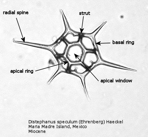
Silicoflagellates
Compound Microscope Labeled Diagram | Quizlet QUESTION. The total magnification of a specimen being viewed with a 10X ocular lens and a 40X objective lens is. 15 answers. QUESTION. a mosquito beats its wings up and down 600 times per second, which you hear as a very annoying 600 Hz sound. if the air outside is 20 C, how far would a sound wave travel between wing beats. 2 answers.

Compound Microscope Parts and Functions | Science fair projects ...
Two-Photon Excitation Microscopy for the Study of Living Cells … The development of miniature two-photon microscope systems and endoscopic or in vivo light delivery has broadened the range of sites than can be accessed. With such miniaturization, a two-photon microscope system can be mounted on freely moving mice, allowing longitudinal imaging studies (Flusberg et al., 2005; Piyawattanametha et al., 2009 ...

Microscope - The Gemology Project
35 Latest Compound Microscope Diagram - Perangkat Sekolah There are three structural parts of the microscope ie. Zoom microscopes have a fixed working distance throughout the zoom range. Parts Of A Microscope With Functions And Labeled Diagram Compounds are used for viewing standard 1 by 3 1mm thick transparent specimen slides with cover slips. 35 latest compound microscope diagram. It also carriers the … 35 Latest Compound Microscope Diagram Read ...

View Product Photos
Compound Microscope Diagram Blank - 16 images - types of microscopes ... microscope diagram labeled unlabeled and blank parts of a microscope. Compound Microscope Diagram Blank. Here are a number of highest rated Compound Microscope Diagram Blank pictures on internet. We identified it from reliable source. Its submitted by presidency in the best field. We put up with this kind of Compound Microscope Diagram Blank ...

Multiple Choice Quiz on Compound Microscope Parts and Functions
Electron microscope - Wikipedia An electron microscope is a microscope that uses a beam of accelerated electrons as a source of illumination. As the wavelength of an electron can be up to 100,000 times shorter than that of visible light photons, electron microscopes have a higher resolving power than light microscopes and can reveal the structure of smaller objects.. Electron microscopes use shaped magnetic …

IBG: IBG 102 LAB REPORT
Parts of a microscope with functions and labeled diagram Q. List down the 18 parts of a Microscope. 1. Ocular Lens (Eye Piece) 2. Diopter Adjustment 3. Head 4. Nose Piece 5. Objective Lens 6. Arm (Carrying Handle) 7. Mechanical Stage 8. Stage Clip 9. Aperture 10. Diaphragm 11. Condenser 12. Coarse Adjustment 13. Fine Adjustment 14. Illuminator (Light Source) 15. Stage Controls 16. Base 17.
The Compound Light Microscope
Microscope Parts and Functions Microscope Parts and Functions With Labeled Diagram and Functions How does a Compound Microscope Work?. Before exploring microscope parts and functions, you should probably understand that the compound light microscope is more complicated than just a microscope with more than one lens.. First, the purpose of a microscope is to magnify a …

Buccal smear | Medical Laboratories
(b) Why both objective and eyepiece of a compound microscope must have ... Question (a) Draw the labelled ray diagram for the formation of image by a compound microscope. Derive an expression for its total magnification (or magnifying power), when the final image is formed at the near point. (b) Why both objective and eyepiece of a compound microscope must have short focal lengths?

Molecular Make Up Of Cells | Cells: The Basic Units Of Life | Siyavula
Draw a labelled diagram of an image formed by a compound microscope ... Question Draw a labelled diagram of an image formed by a compound microscope, with the image at least distance of distinct vision. Write any one expression for its magnifying power. Medium Solution Verified by Toppr Expression of magnifying power of a compound microscope is given by: m=− u o v o (1+ f e D ) Where v o
Post a Comment for "39 labeled diagram of compound microscope"