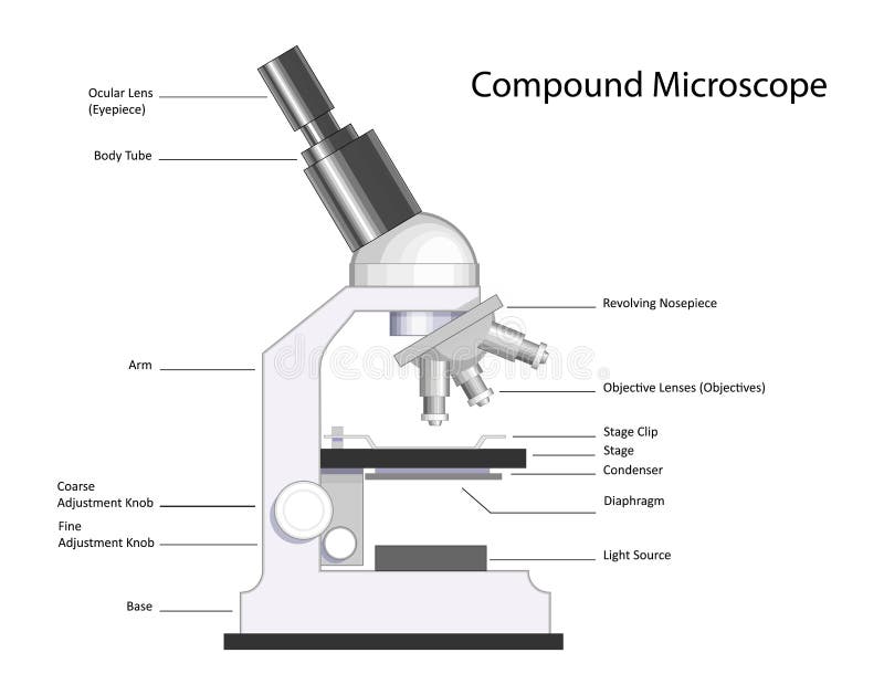
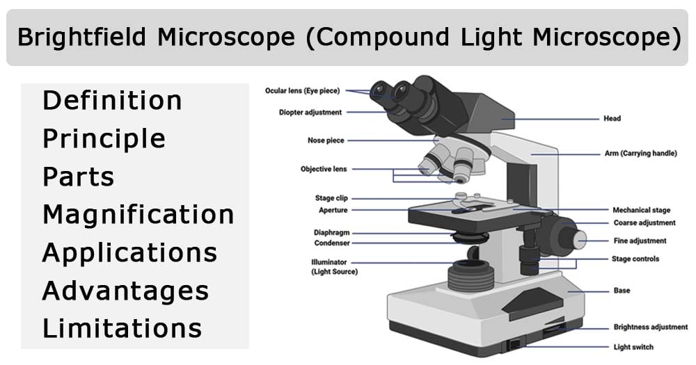
38 compound light microscope labelled
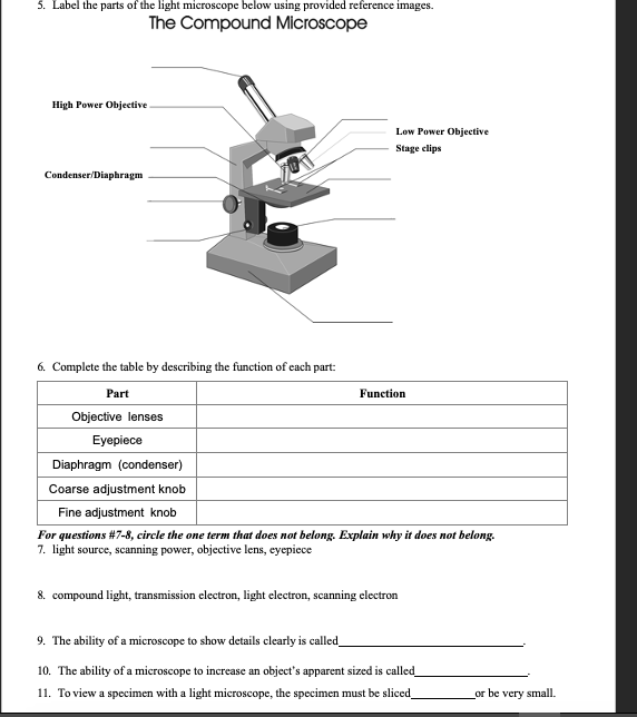
PDF The Compound Light Microscope - teachers.wrdsb.ca The Compound Light Microscope TASK Refer to page 605 in your text to: 1. Name each of the structures described in the table to the right. 2. Match each structure to the letter in the diagram below. ** ALWAYS USE TWO HANDS TO CARRY A MICROSCOPE** Letter Structure Function joins body tube to base supports the entire microscope compound-light-microscope-labeled-compound-light-microscope-parts-and ... View compound-light-microscope-labeled-compound-light-microscope-parts-and-functions-worksheet-www-within from BIOL 120 at Cerritos College. let's Review! 9. Eyepiece 1 . Body tube 2. Nosepiece 10.
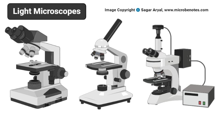
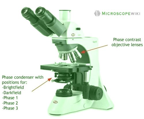
Compound Light Microscope Optics, Magnification and Uses AmScope 40X-1000X LED Cordless Student Compound Microscope. AmScope 40X-2500X LED Digital Binocular Compound Microscope with 3D Stage and 5MP USB Camera. Omax 40X-2000X Compound Biological Microscope with built-in Camera Omax 40X-2000X Lab LED Binocular Compound Microscope Promotion Set
Compound light microscope labelled
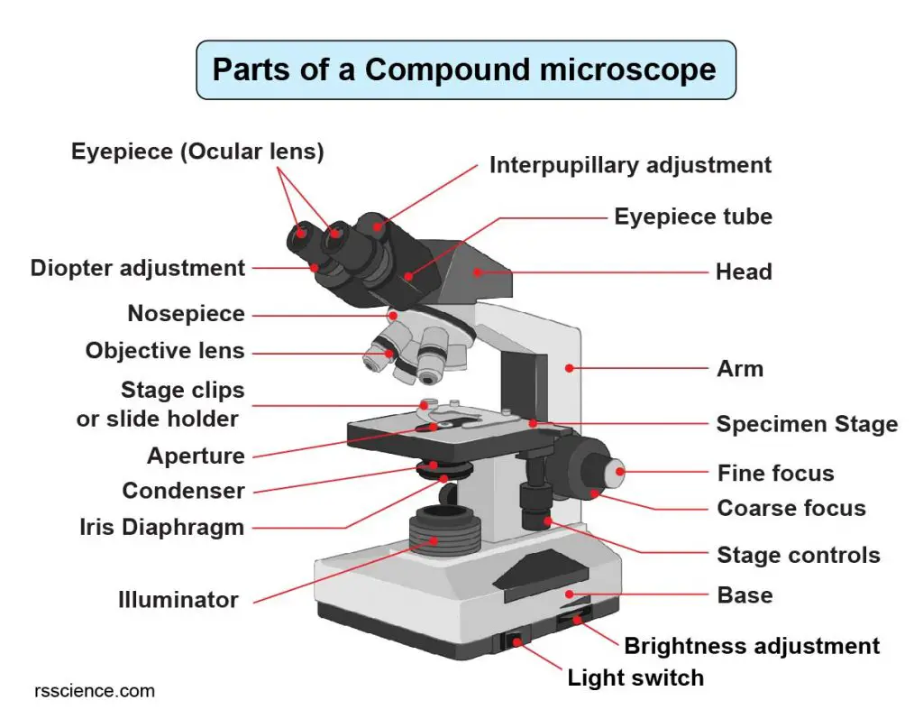
A general design of caging-group-free photoactivatable ... Jul 21, 2022 · a–c, Confocal imaging of U2OS cells labelled with Abberior LIVE 560 tubulin (AL-560, 500 nM) and vimentin filaments labelled with compound 21 (100 nM) before (a) and after (b) photobleaching of ... Alwaysales - always your best choice Light, Airy, and Silky to Touch - Perfect Way to Treat Yourself Genius Double-Chamber Ice Cube Maker Holds Up to 120 Cubes, Essentially Replaces 10 of Normal Trays Compound Microscope Parts, Functions, and Labeled Diagram The individual parts of a compound microscope can vary heavily depending on the configuration & applications that the scope is being used for. Common compound microscope parts include: Compound Microscope Definitions for Labels Eyepiece (ocular lens) with or without Pointer: The part that is looked through at the top of the compound microscope. Eyepieces typically have a magnification between 5x & 30x.
Compound light microscope labelled. Copper-mediated radioiodination and radiobromination via aryl ... The labeled compound was identified by UV- and radio-HPLC analysis of a standard sample and the synthesized radiolabeled compound. 4.5. In vitro stability study [125 I]mIB-PS and [77 Br]mBrB-PS were synthesized using method II. The isolated [125 I]mIB-PS or [77 Br]mBrB-PS was dissolved in water and was trapped in Sep-Pack light C 18 (Waters ... A Study of the Microscope and its Functions With a Labeled Diagram ... To better understand the structure and function of a microscope, we need to take a look at the labeled microscope diagrams of the compound and electron microscope. These diagrams clearly explain the functioning of the microscopes along with their respective parts. Man's curiosity has led to great inventions. The microscope is one of them. A general design of caging-group-free photoactivatable ... - Nature 21.07.2022 · U2OS cells stably expressing a vimentin–HaloTag fusion construct 48 were labelled with compound 21 and imaged with a confocal microscope equipped with a subpicosecond pulsed laser (Fig. 3c). Microscope Types (with labeled diagrams) and Functions A compound microscope: Is used to view samples that are not visible to the naked eye Uses two types of lenses - Objective and ocular lenses Has a higher level of magnification - Typically up to 2000x Is used in hospitals and forensic labs by scientists, biologists and researchers to study micro organisms Compound microscope labeled diagram
Parts of a microscope with functions and labeled diagram - Microbe Notes Head - This is also known as the body. It carries the optical parts in the upper part of the microscope. Base - It acts as microscopes support. It also carries microscopic illuminators. Arms - This is the part connecting the base and to the head and the eyepiece tube to the base of the microscope. Compound Microscope - Diagram (Parts labelled), Principle and Uses Also called as binocular microscope or compound light microscope, it is a remarkable magnification tool that employs a combination of lenses to magnify the image of a sample that is not visible to the naked eye. Compound microscopes find most use in cases where the magnification required is of the higher order (40 - 1000x). What is a Compound Microscope? - Study.com The body of the compound light microscope is the main part of the microscope, not to include the lights, focusing block, or stand of the microscope. The objective lenses and eyepiece are a part of ... Microscope Parts, Function, & Labeled Diagram - slidingmotion Condenser. The condenser is to focus the light, which passes from the microscopic illuminator to the specimen. This condenser is located just below the diaphragm. This diaphragm is one of the important parts of the compound microscope which will help to get an accurate and sharp image. The condenser has a magnification power of 400X and above.
Correlating ligand-to-metal charge transfer with voltage ... Sep 16, 2021 · The electrochemical behaviour of LTFO was tested versus Li between 2.0 and 4.8 V at a C/10 rate (Fig. 1b).On oxidation, the removal of Li occurs via a single-step plateau located at ~4.25 V as ... Laboratory Information System (LIS) - HEALTHCARE SERVICE … 06.09.2014 · Date First Published: September 6, 2014Date Last Revised: March 20, 2020 The name Laboratory Information System is more appropriate than say Pathology information system, since the Pathology department serves many functions, and systems should be designed to be functional rather than departmental. Clinical functions such as clinical microbiology … Electron microscope - Wikipedia An electron microscope is a microscope that uses a beam of accelerated electrons as a source of illumination. As the wavelength of an electron can be up to 100,000 times shorter than that of visible light photons, electron microscopes have a higher resolving power than light microscopes and can reveal the structure of smaller objects.. Electron microscopes use shaped magnetic … Compound Light Microscope Labeling - Printable - PurposeGames.com About this Worksheet. This is a free printable worksheet in PDF format and holds a printable version of the quiz Compound Light Microscope Labeling. By printing out this quiz and taking it with pen and paper creates for a good variation to only playing it online.
Compound Microscope: Parts of Compound Microscope - BYJUS (A) Mechanical Parts of a Compound Microscope. 1. Foot or base. It is a U-shaped structure and supports the entire weight of the compound microscope. 2. Pillar. It is a vertical projection. This stands by resting on the base and supports the stage. 3. Arm. The entire microscope is handled by a strong and curved structure known as the arm. 4. Stage
Labelled Diagram of Compound Microscope - Biology Discussion The below mentioned article provides a labelled diagram of compound microscope. Part # 1. The Stand: The stand is made up of a heavy foot which carries a curved inclinable limb or arm bearing the body tube. The foot is generally horse shoe-shaped structure (Fig. 2) which rests on table top or any other surface on which the microscope in kept.
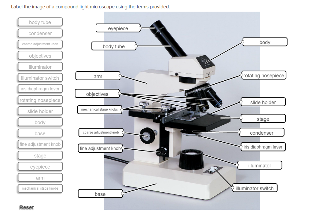
Solved Label the image of a compound light microscope using - Chegg Expert Answer. 100% (17 ratings) Transcribed image text: Label the image of a compound light microscope using the terms provided.
Parts of the Microscope with Labeling (also Free Printouts) Image 2: The body tube part of a microscope is where the ray of light is bent to allow the object being viewed to enlarge by the scope. Picture Source: slideplayer.com 3. Turret/Nose piece. It is the revolving part of the microscope. It allows the use of different types of objective lenses by simply rotating the top part of the turret.
Compound Microscope Parts - Labeled Diagram and their Functions What is a "compound microscope"? A compound microscope is the most common type of light (optical) microscopes. The term "compound" refers to the microscope having more than one lens. Basically, compound microscopes generate magnified images through an aligned pair of the objective lens and the ocular lens.
Microscope Parts and Functions Microscope Parts and Functions With Labeled Diagram and Functions How does a Compound Microscope Work?. Before exploring microscope parts and functions, you should probably understand that the compound light microscope is more complicated than just a microscope with more than one lens.. First, the purpose of a microscope is to magnify a small object or to magnify the fine details of a larger ...
Label the microscope — Science Learning Hub All microscopes share features in common. In this interactive, you can label the different parts of a microscope. Use this with the Microscope parts activity to help students identify and label the main parts of a microscope and then describe their functions. Drag and drop the text labels onto the microscope diagram. If you want to redo an answer, click on the box and the answer will go back to the top so you can move it to another box.
Label the Parts of a Compound Light microscope Label the Parts of a Compound Light microscope. Biology Junction Team April 21, 2017 Uncategorized.
Read Important Questions Class 12 Physics of Chapter 9 1 Mark Questions. 1. A person standing before a concave mirror cannot see his image unless he is beyond the centre of curvature. Why? Ans: Let a man stand beyond focus i.e., between focus and centre of curvature, then the image formed will be real and inverted and is formed beyond C (beyond him). Thus, he cannot see the image.
Ark Elvin Academy Year 9 Science Study Pack Autumn assessment … • A light microscope shines a beam of light across a thin, dead, stained specimen. • The resolution (ability to distinguish between two points) and magnification of a light microscope is high enough the view the nucleus and cell membrane. • Most organelles are too small to be viewed with a light microscope. 5. Diffusion Key information:
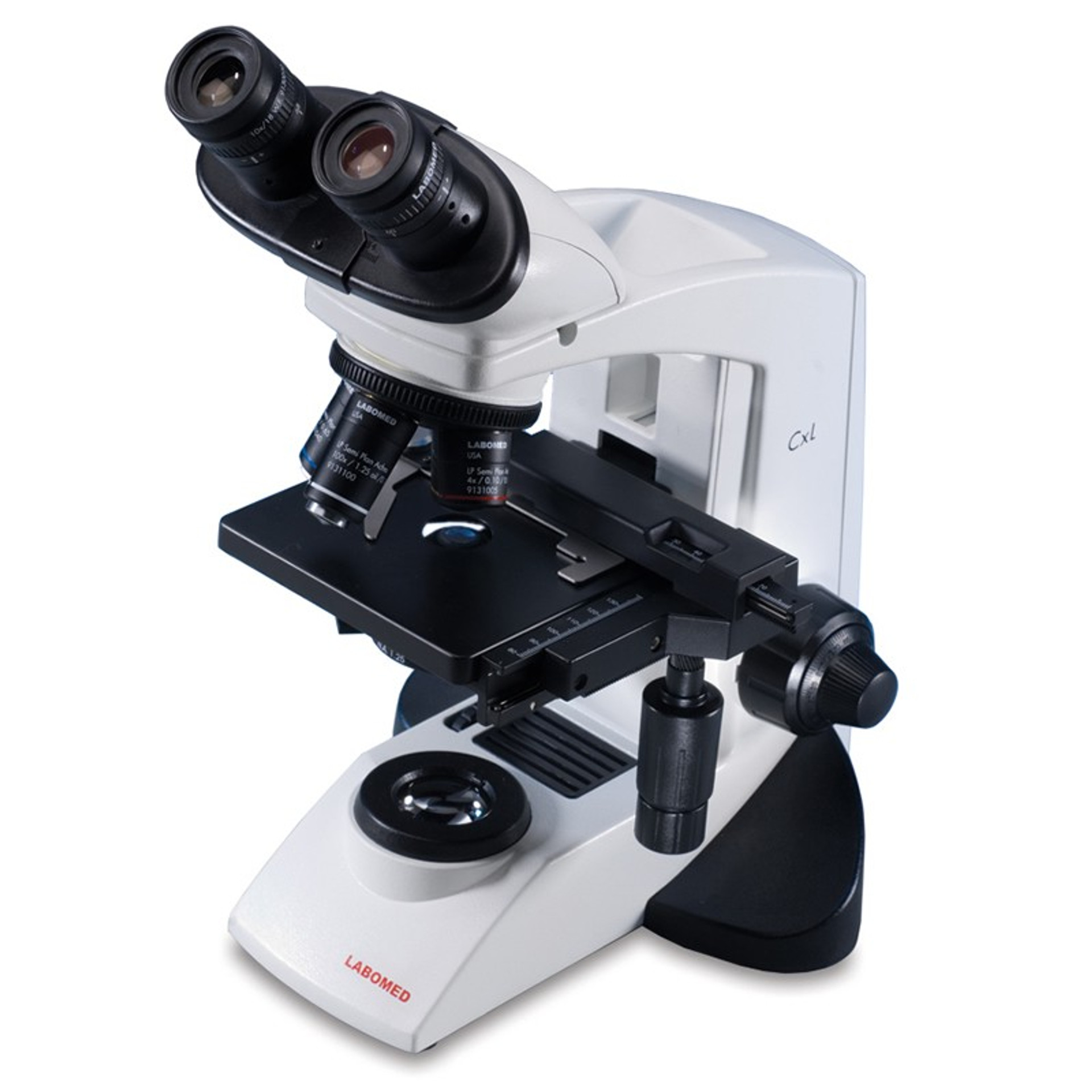
10 Best Compound Microscopes (Summer 2022) - The Complete Guide The OMAX 40X-2500X LED Binocular Compound Lab Microscope is our top choice for a compound microscope. It is an award-winning device that comes with a swiveling head, revolving nosepiece, integrated illumination, and a generous warranty.
Fluorescence Microscopy - Explanation and Labelled Images 16.12.2020 · A fluorescence microscope uses a higher intensity light to illuminate the samples. Parts of a Fluorescence Microscope A powerful light source (xenon or mercury arch lamp) : The light emitted from the mercury arc lamp is 10-100 times brighter than most incandescent lamps and provides light in a wide range of wavelengths, from ultra-violet to the infrared.
Compound Light Microscope: Everything You Need to Know A compound microscope is a type of light microscope that uses a compound lens system to magnify specimens for up to 1000x or more. It's made up of at least one objective lens and at least one ocular lens, as well as a light source, condenser, and other essential parts.
Label the Microscope - Pinterest Label the Microscope Description Worksheet identifying the parts of the compound light microscope. Answer key: 1. Body tube 2. Revolving nosepiece 3. Low power objective 4. Medium power objective 5. High power objective 6. Stage clips 7. Diaphragm 8. Light source 9. Eyepiece 10. Arm 11. Stage 12. Coarse adjustment knob 13. Fine adjustment knob 14.
Parts of a Compound Microscope and Their Functions - NotesHippo Compound microscope mechanical parts (Microscope Diagram: 2) include base or foot, pillar, arm, inclination joint, stage, clips, diaphragm, body tube, nose piece, coarse adjustment knob and fine adjustment knob. Base: It's the horseshoe-shaped base structure of microscope. All of the other components of the compound microscope are supported ...
Copper-mediated radioiodination and radiobromination via aryl … We recently reported that copper-mediated radioiodination via boronic precursors is effective for the direct labeling of peptides. 12 This reaction does not require heating and selectively provides a desired radiolabeled compound. 13 Thus, we expect this to be an effective method for synthesizing PSMA imaging probes. Moreover, a copper-mediated radiobromination method of …
Compound Microscope- Definition, Labeled Diagram, Principle, Parts, Uses The illuminator is the light source for a microscope. A compound light microscope mostly uses a low voltage bulb as an illuminator. The stage is the flat platform where the slide is placed. Nosepiece and Aperture. Nosepiece is a rotating turret that holds the objective lenses. The viewer spins the nosepiece to select different objective lenses.
PDF Parts of the Light Microscope - Science Spot Supports the MICROSCOPE D. STAGE CLIPS HOLD the slide in place C. OBJECTIVE LENSES Magnification ranges from 10 X to 40 X F. LIGHT SOURCE Projects light UPWARDS through the diaphragm, the SPECIMEN, and the LENSES H. DIAPHRAGM Regulates the amount of LIGHT on the specimen E. STAGE Supports the SLIDE being viewed K. ARM Used to SUPPORT the
Parts of the Compound Microscope - Houston Community College Parts of the Compound Microscope Use Figure 2 as a guide to locate the major parts of the compound microscope. a. Base: The bottom, flat part that supports the microscope. b. Arm: The straight or curved vertical part that connects the base to the upper portion. c. Body Tube: Extends from the arm and contains the ocular lens and the rotating
Fluorescence - Wikipedia Fluorescence is the emission of light by a substance that has absorbed light or other electromagnetic radiation.It is a form of luminescence.In most cases, the emitted light has a longer wavelength, and therefore a lower photon energy, than the absorbed radiation.
Why Is The Light Microscope Called A Compound Microscope? The light microscope is sometimes called a compound microscope because of its ability to use several lenses at a time, just like a compound microscope. Normal light microscopes cannot reach the highest magnifications because of their single-lens magnification system.
State of the Art in Defect Detection Based on Machine Vision May 26, 2021 · White light source is a multi-wavelength compound light, which is widely used. High brightness white light source is suitable for color image shooting. The wavelength of blue light is between 430 and 480 nm, and is suitable for sheet metal, machining parts, and other products with a silver colored background, as well as metal printing on film.
Eye - Wikipedia Eyes are organs of the visual system.They provide living organisms with vision, the ability to receive and process visual detail, as well as enabling several photo response functions that are independent of vision.Eyes detect light and convert it into electro-chemical impulses in neurons.In higher organisms, the eye is a complex optical system which collects light from the …
Correlating ligand-to-metal charge transfer with voltage ... - Nature 16.09.2021 · The use of anionic redox chemistry in high-capacity Li-rich cathodes is being hampered by voltage hysteresis, the origin of which remains obscure. Now it has been shown that sluggish ligand-to ...
An Overview of Disintegrants | LFA Tablet Presses 1. Rapid dissolution increases the rate of absorption of the active ingredient by the body, producing the desired therapeutic action. Note that tablets that are labelled as chewable generally do not require a disintegrant to be incorporated in the formulation. Tablet Disintegrants Methods. Tablets disintegrate by: capillary action and wicking
Eye - Wikipedia Photoreception is phylogenetically very old, with various theories of phylogenesis. The common origin of all animal eyes is now widely accepted as fact.This is based upon the shared genetic features of all eyes; that is, all modern eyes, varied as they are, have their origins in a proto-eye believed to have evolved some 650-600 million years ago, and the PAX6 gene is considered a key factor in ...
Compound Microscope Labeled Diagram | Quizlet QUESTION. The total magnification of a specimen being viewed with a 10X ocular lens and a 40X objective lens is. 15 answers. QUESTION. a mosquito beats its wings up and down 600 times per second, which you hear as a very annoying 600 Hz sound. if the air outside is 20 C, how far would a sound wave travel between wing beats. 2 answers.
Compound Microscope: Definition, Diagram, Parts, Uses, Working ... - BYJUS A compound microscope is defined as. A microscope with a high resolution and uses two sets of lenses providing a 2-dimensional image of the sample. The term compound refers to the usage of more than one lens in the microscope. Also, the compound microscope is one of the types of optical microscopes. The other type of optical microscope is a simple microscope.
Compound Microscope Parts, Functions, and Labeled Diagram The individual parts of a compound microscope can vary heavily depending on the configuration & applications that the scope is being used for. Common compound microscope parts include: Compound Microscope Definitions for Labels Eyepiece (ocular lens) with or without Pointer: The part that is looked through at the top of the compound microscope. Eyepieces typically have a magnification between 5x & 30x.
Alwaysales - always your best choice Light, Airy, and Silky to Touch - Perfect Way to Treat Yourself Genius Double-Chamber Ice Cube Maker Holds Up to 120 Cubes, Essentially Replaces 10 of Normal Trays
A general design of caging-group-free photoactivatable ... Jul 21, 2022 · a–c, Confocal imaging of U2OS cells labelled with Abberior LIVE 560 tubulin (AL-560, 500 nM) and vimentin filaments labelled with compound 21 (100 nM) before (a) and after (b) photobleaching of ...















Post a Comment for "38 compound light microscope labelled"