39 labeled histology slides
Uterus histology slide with labeled diagram and identification points Uterus histology slide labeled diagrams. In the uterus histology slide diagram, you will find all the important features. It shows the three different layers of the uterus along with the lining epithelium, uterine glands, and smooth muscle layers. Suggested histology slides for you - The uterine tube histology slide with a labeled diagram Histology Slides 1 - Loyola University Chicago Slide 15 EM of liver cell (hepatocyte), showing aggregates of glycogen (1). Compare their size with the ribosomes lining the endoplasmic reticulum (2). Gallbladder Slide 16 Wall of gall bladder, showing high, branching mucosal folds. These are not villi. The rest of the wall contains connective tissue and thin strands of smooth muscle. Slide 17
HistologySlide - Identification of Microscopic Slides HistologySlide - Identification of Microscopic Slides Histology Slide Identification with Identifying Characteristics Collection of microscopic images, labeled diagrams, videos, and more Latest from HistologySlide Slide Identifying Points Slide Identifying Points Slide Identifying Points Slide Identifying Points Slide Identifying Points

Labeled histology slides
PPT Histology Slides - Oakton Histology Slides Biology 131 - Anatomy and Physiology Instructor: Ruth Williams Epithelial Tissues No intercellular matrix. Avascular Contains nerve endings Lie on a basement membrane. Able to undergo mitosis. Develop from all three fetal tissues. One surface of cells is exposed to a space or cavity. Epididymis Histology Slide and Identification Points with Labeled ... Epididymis Histology Slide and Identification Points with Labeled Diagram 19/11/2021 by anatomylearner The epididymis is the comma-shaped structure in the male organ system that divides into head, body, and tail. It is made of highly coiled, tortuous ductus tubules and vascular connective tissue. Histology guide: Definition and slides | Kenhub At a histological level, both the heart and blood vessels consist of three layers: Endothelial layer - epithelial tissue formed by simple squamous (endothelial) cells. In the heart, this layer is referred to as endocardium. Muscular layer - smooth muscle in the blood vessels, cardiac muscle (myocardium) in the heart.
Labeled histology slides. Microscope Slides of Cells and Tissues | Histology Guide This virtual slide box contains 275 microscope slides for the learning histology. Fig 023 Types of Tissue Cells and Tissues Tissues are classified into four basic types: epithelium, connective tissue (includes cartilage, bone and blood), muscle, and nervous tissue. Chapter 1 The Cell Chapter 2 Epithelium Chapter 3 Connective Tissue Chapter 4 Muscle Histology Slides - Napa Valley College BIOL 218 Human Anatomy; BIOL 219 Human Physiology; BIOL 240 General Zoology; People Sites > Dan Clemens > Histology Slides > > People ... Histology Slides Clemens BIOL 218 Histology Slide Images Exercise 2 - Cells 02_Blood_100X 02_Blood_400X 02_Neurons ... Colon Histology Slide with Labeled Diagram - AnatomyLearner Colon Histology Slide with Labeled Diagram 04/06/2022 04/06/2022 by anatomylearner The colon histology slide possesses the typical four layers of a tubular organ - mucosa, submucosa, muscularis, and serosa. But, there are no permanent plica circularis and villi in the colon slide as found in the different segments of the small intestine. Ileum Histology Slides Labeled - histology digestion lab ileum, ileum ... Ileum Histology Slides Labeled - 16 images - histology digestion lab ileum, 2nd year mbbs histology slides medical ebooks and articles, histology slides database ileum histology slides, bgdb practical upper gastrointestinal tract histology embryology,
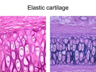
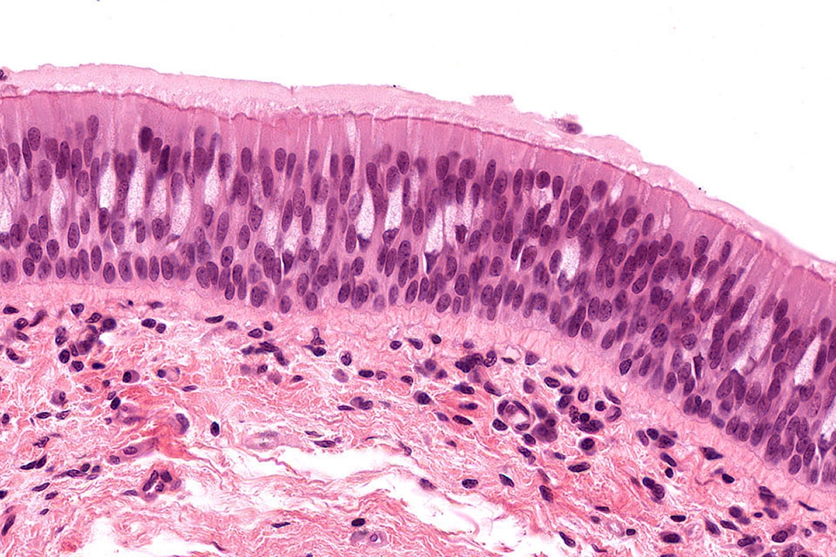
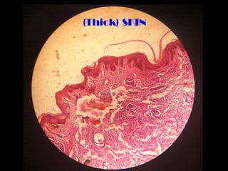
Skin Histology Slide Identification - AnatomyLearner I would like to show you the different histological features from both thick and thin skin histology slides with a labeled diagram. I hope these skin microscope slide labeled diagrams might help you to identify and learn all the structures. If you need more skin microscope slide labeled diagram, please follow anatomy learner on social media. I ... Slides - HistologySlide 06/11/2021 by histologyslide Adrenal gland histology slide diagram and identification points The adrenal gland consists of a yellow peripheral region (the cortex), and a brown central region, the medulla. Here, you will learn all the features from the adrenal gland histology slide with a labeled diagram and perfect identification points. Ileum Histology Slide with Labeled Diagram and Identification Points The wall of the ileum histology consists of four distinguished layers - tunica mucosa, submucosa, muscular, and serosa. You will find thin and slender villi on the tunica mucosa layer of the ileum slide. The simple columnar epithelium lines these villi of the ileum. So, you will find the intervillous spaces between the villi of the ileum structure. Histology Slides Identification from Different Organ Systems This article will show you histology slides from the following different organs system of an animal's body with identifying features. #1. Histology slide of epithelial tissue #2. General connective tissue histology slide #3. Histology slides of special connective tissue (blood, bone, and cartilage) #4. Muscular tissue histology slide #5.
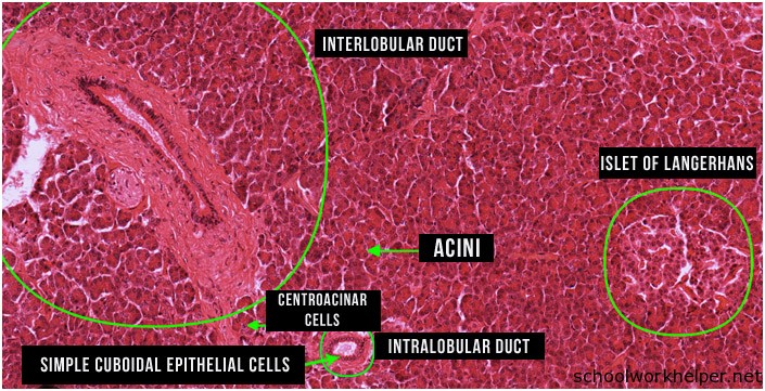
Virtual Slide List | histology It will also benefit the publication of several new topics (Hematology, Pathogen ID, and Gross Anatomy). Any size contribution is welcomed and will help us to provide these popular review tools to students at the University of Michigan and to many more worldwide. ... Virtual Slide List for Histology Course . Slide Category Cecum Histology Slide with Labeled Image and Diagram Generally, the tunica muscularis layer of the cecum histology slide consists of a thick inner circular and thin outer longitudinal smooth muscle layer. The outer longitudinal smooth muscle layer forms large, flat, smooth muscle bands in pig and horse cecum. This smooth muscle band of the pig and horse cecum is known as the taenia Ceci. Histology: Labelled Slides | SchoolWorkHelper Histology: Labelled Slides Aorta Basophils Cardiac Muscle Cardiac Muscle Longtudinal Cerebellar Cortex Cerebral Cortex Spinal Cord Eosinophils Epiphyseal plate Femoral Artery Femoral Vein Howship's lacunae Jejunum lamina propria Liver Lymph node Lymphocyte Monocyte Spinal cord (silver) Neutrophil Pacinian corpuscle Peripheral Nerve Ovary histology slide diagram and identification points In the ovary histology slide labeled diagram, you will find all the features that are mentioned above. Identification of other slides from the female organs Uterine tube histology slide with its identification points Uterus histology slide with its identification points Mammary glands histology slide with its identification points
Histology Guide - virtual microscopy laboratory Histology is the study of the microanatomy of cells, tissues, and organs as seen through a microscope. It examines the correlation between structure and function. Histology Guide teaches the visual art of recognizing the structure of cells and tissues and understanding how this is determined by their function.
Blood Histology Slides with Description and Labeled Diagram In the blood histology slide (smear), all of the cells mentioned earlier with their useful identifying features. There are five different types of leukocytes in blood - neutrophils, eosinophils, basophils, monocytes, and lymphocytes. The neutrophils, eosinophils, and basophils are known as the polymorphonuclear granulocytes.
Slides of Histology | Anatomy and Physiology I - Lumen Learning Slides of Histology Learning Objectives Be able to describe the functions of cells commonly found in connective tissue and identify them. Be able to recognize interstitial (fibrillar) collagens and elastic fibers at the light and electron microscopic levels.
Histology guide: Definition and slides | Kenhub At a histological level, both the heart and blood vessels consist of three layers: Endothelial layer - epithelial tissue formed by simple squamous (endothelial) cells. In the heart, this layer is referred to as endocardium. Muscular layer - smooth muscle in the blood vessels, cardiac muscle (myocardium) in the heart.
Epididymis Histology Slide and Identification Points with Labeled ... Epididymis Histology Slide and Identification Points with Labeled Diagram 19/11/2021 by anatomylearner The epididymis is the comma-shaped structure in the male organ system that divides into head, body, and tail. It is made of highly coiled, tortuous ductus tubules and vascular connective tissue.
PPT Histology Slides - Oakton Histology Slides Biology 131 - Anatomy and Physiology Instructor: Ruth Williams Epithelial Tissues No intercellular matrix. Avascular Contains nerve endings Lie on a basement membrane. Able to undergo mitosis. Develop from all three fetal tissues. One surface of cells is exposed to a space or cavity.





![PDF] Use of Labeled Histology Images with Key Identification ...](https://d3i71xaburhd42.cloudfront.net/9e03ebd5b9681a62269a4d60cd6b0af01715e6ca/2-Figure1-1.png)




Post a Comment for "39 labeled histology slides"