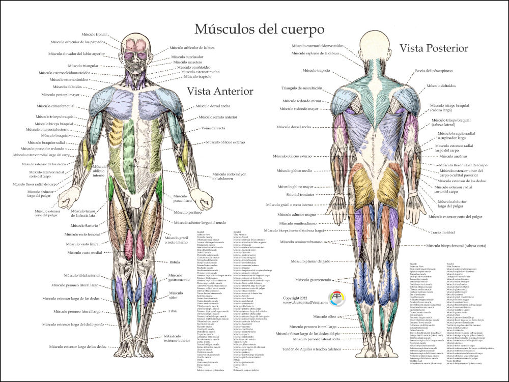
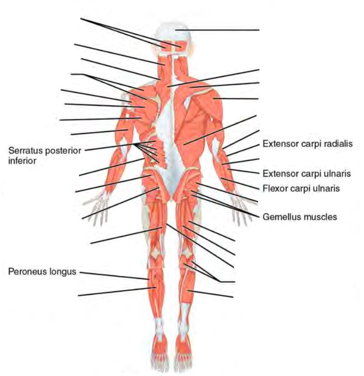
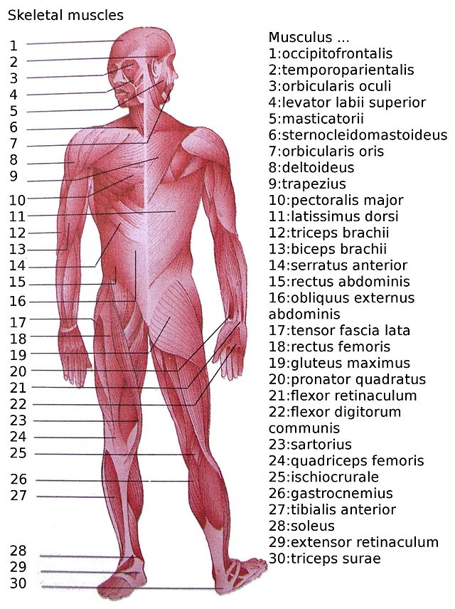
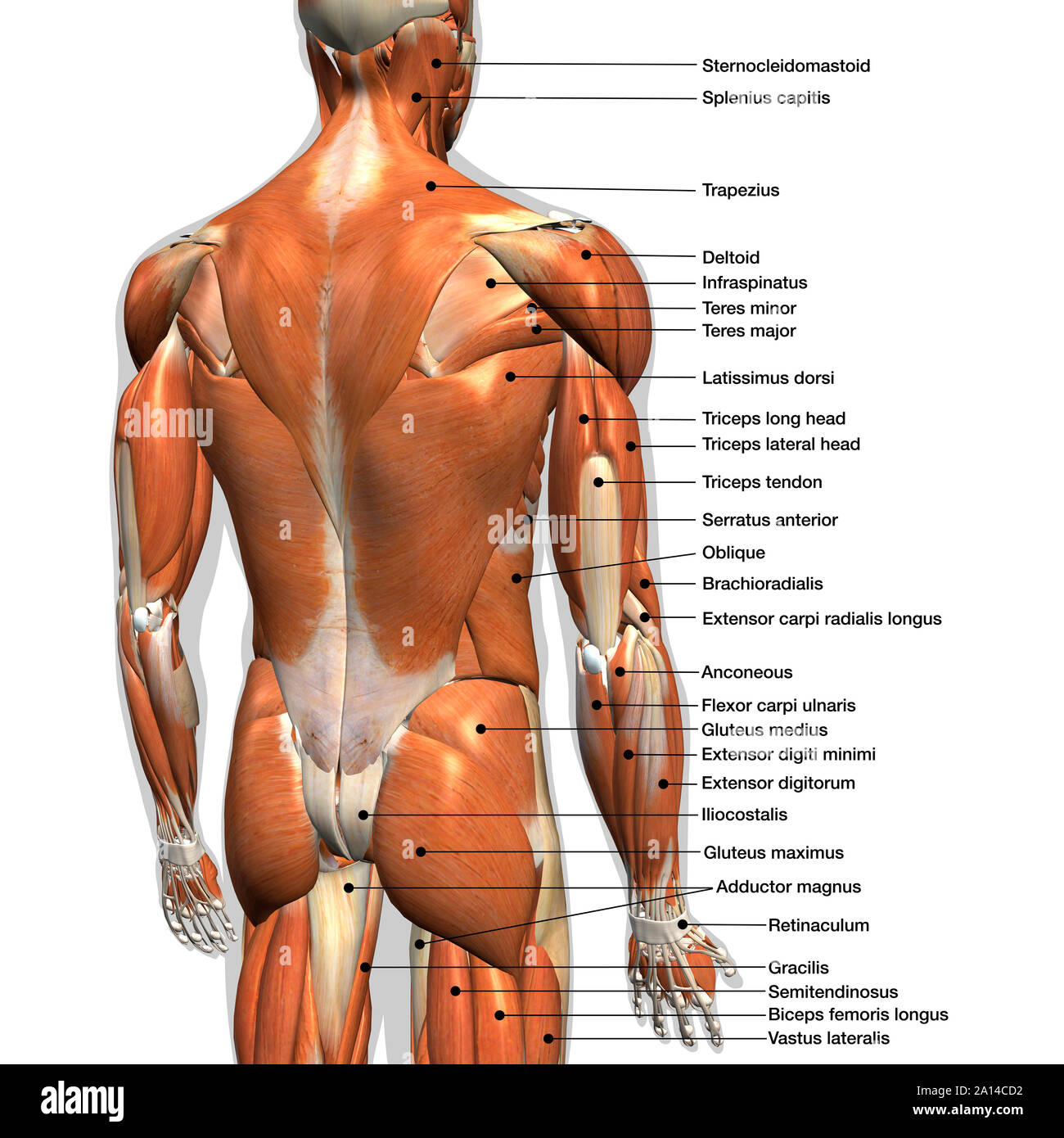
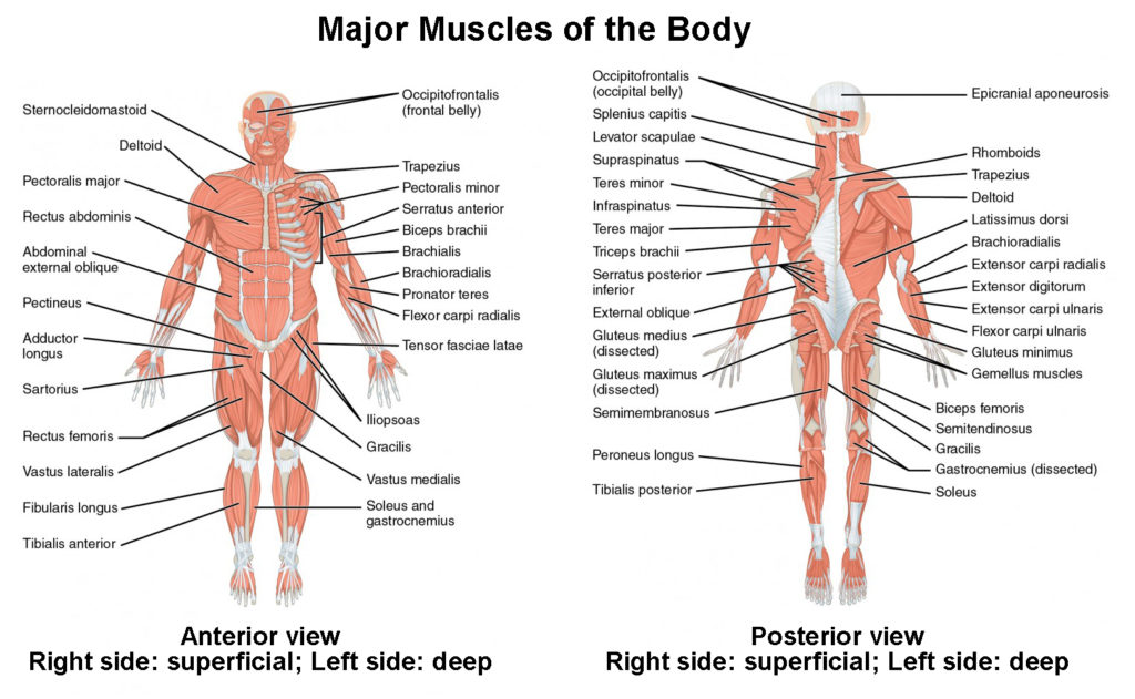
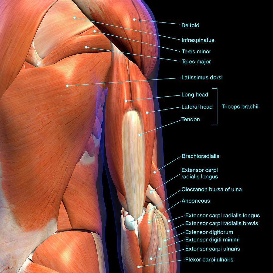
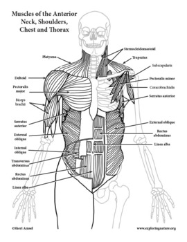
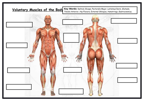
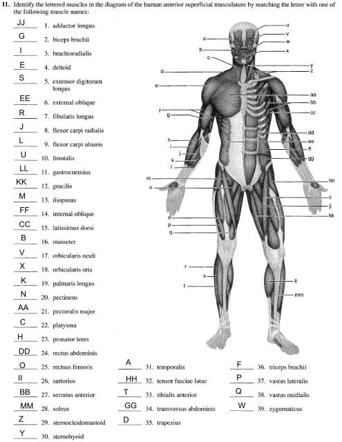
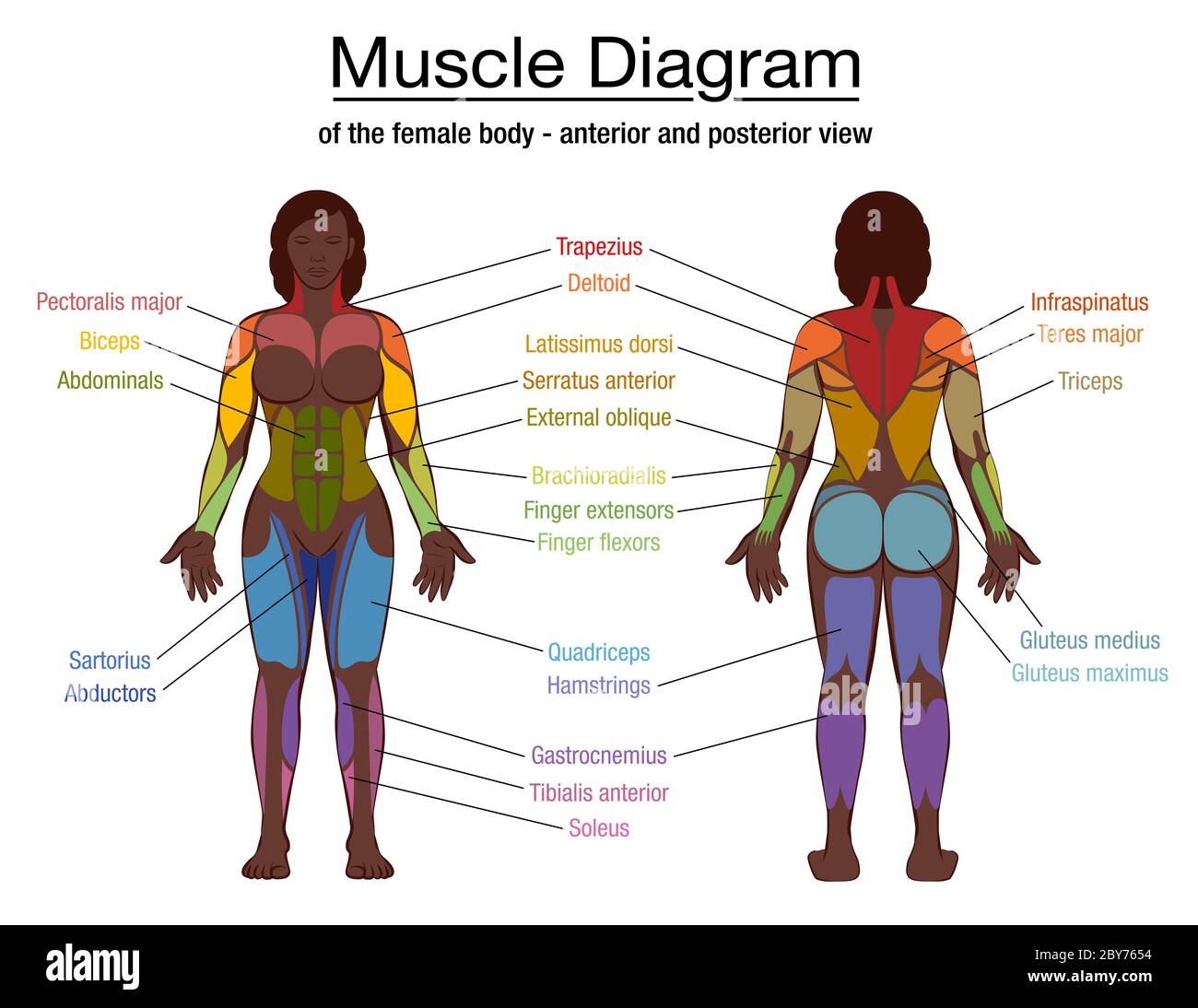
41 labeled muscle diagram
Learn all muscles with quizzes and labeled diagrams | Kenhub Labeled diagram View the muscles of the upper and lower extremity in the diagrams below. Use the location, shape and surrounding structures to help you memorize each muscle. Once you're feeling confident, it's time to test yourself. Unlabeled diagram See if you can label the muscles yourself on the worksheet available for download below. Muscle anatomy reference charts: Free PDF download | Kenhub This muscle chart eBook covers the following regions: Inner hip & gluteal muscles Anterior, medical and posterior thigh muscles Anterior, lateral and posterior leg muscles Dorsal and plantar foot muscles This eBook contains high-quality illustrations and validated information about each muscle. It is available for free. Download free PDF (8.5MB)
Labelled Diagram Of The Muscles In The Human Body - Anatomy Note The muscles of the human body can be categorized into a number of groups which include muscles relating to the head and neck, muscles of the torso or trunk, muscles of the upper limbs, and muscles of the lower limbs. The human body has three different types of muscles. They include: skeletal muscles, smooth muscles, cardiac muscles.
Labeled muscle diagram
PDF ANATOMY OF THE MUSCULAR SYSTEM - Midland Independent School District individual muscle fibers, (b) surrounds groups of skeletal muscle fibers (fascicles), and (c) covers the muscle as a whole. 2. Name the tough connective tissue cord that serves to attach a muscle to a bone. 3. Name three types of fiber arrangements seen in skeletal muscle. Foot Diagram: Labeled Anatomy | Science Trends Jul 05, 2018 · The foot diagram has a complex structure made up of bones, ligaments, muscles, and tendons. Understanding the structure of the foot is best done by looking at a foot diagram where the anatomy has been labeled. If you would like to learn all the parts of the foot structure, you have come to the right place. shin muscles diagram muscular system muscles anatomy physiology leg nurseslabs lower muscle diagram body foot calf gastrocnemius human joint. Running Shin Splints Treated Graston Technique Sioux City chiropractor-sioux-city.com. shin leg lower splints muscles labeled anatomy muscle tibialis knee anterior tibia tendons tissue bone sioux chiropractor helena graston pain
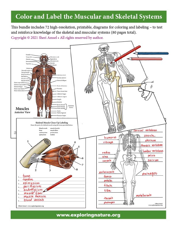
Labeled muscle diagram. Printable Human Skeleton Diagram – Labeled, Unlabeled, and ... This diagram comes in six versions, all combined into one PDF: 1. Human Skeleton Diagram – Color – Labeled 2. Human Skeleton Diagram – Color – Blanks to Fill in 3. Human Skeleton Diagram – Color -No Labels or Blanks (For bulletin boards/decorations etc) 4. Human Skeleton Diagram – Black and White – Labeled 5. labeled muscle diagram diagram muscle muscular muscles human system anatomy medical The Labeled Diagram Of The Heart And Blood Flow Blood Coming Out Of medicinebtg.com blood flow heart diagram through ventricle left pathway labeled aorta coming shutterstock medicinebtg Labeled Muscle Diagram Chart Free Download Skeletal System - Labeled Diagrams of the Human Skeleton - Innerbody The skeletal system includes all of the bones and joints in the body. Each bone is a complex living organ that is made up of many cells, protein fibers, and minerals. The skeleton acts as a scaffold by providing support and protection for the soft tissues that make up the rest of the body. The skeletal system also provides attachment points for ... Muscular System Diagram - SmartDraw Create healthcare diagrams like this example called Muscular System Diagram in minutes with SmartDraw. SmartDraw includes 1000s of professional healthcare and anatomy chart templates that you can modify and make your own. 1/29 EXAMPLES EDIT THIS EXAMPLE Text in this Example: Superficial Anterior Muscles Deltoid Frontalis Pectoralis Biceps
Muscular System - Muscles of the Human Body - Innerbody Zygomaticus Major Muscle Zygomaticus Minor Muscle CHEST AND UPPER BACK Abdominal Head of Pectoralis Major Muscle Clavicular Head of Pectoralis Major Muscle Infraspinatus Muscle Latissimus Dorsi Muscle Levator Scapulae Muscle Serratus Anterior Muscle Sternocostal Head of Pectoralis Major Muscle Sternohyoid Muscle Teres Major Muscle Muscle Anatomy: Diagram, Skeletal Muscles, Free Fitness, 2022. Muscles of Hamstring and Back of the Leg (hamstring, gastrocnemius, gluteus maximus) Muscles in the Human Body (Pectoralis Major, Abdominals, Obliques) Muscles of the Upper Limb (Deltoid, Biceps, Forearms) Muscles of Back (Trapezius, Latissimus Dorsi) Muscles in the Arm (triceps, rear deltoid) Muscles of the Hip & Thigh (quadriceps, hips) Cat Skeleton Anatomy with Labeled Diagram - AnatomyLearner May 29, 2021 · Cat skeleton anatomy labeled diagram. Now, I will show you all the bones from the cat skeleton with a diagram. If you find any mistakes in this cat anatomy labeled diagram, please let me know. I hope this cat skeletal system anatomy labeled diagram might help you understand and identify all the cat’s bones. diagram of major muscles Fat Loss, Building Muscle & Staying Fit: Human Anatomy Diagram exercisenz.blogspot.com. anatomy human diagram muscle body organs female skeleton chart physiology muscular muscles bing rear diagrams names drawing fat system study. Muscle Diagrams . leg muscles names 101diagrams anatomia posterior. Muscles - Kids Discover ...
Muscles Body Diagram Illustrations & Vectors - Dreamstime Download 602 Muscles Body Diagram Stock Illustrations, Vectors & Clipart for FREE or amazingly low rates! New users enjoy 60% OFF. 191,356,969 stock photos online. ... Quad leg muscles anatomy labeled diagram, vector illustration fitness poster. Sports physiotherapy educational information. Healthy muscular structure and Dog Skeleton Anatomy with Labeled Diagram - The Place to ... Dec 31, 2021 · In addition, in the diagram, you will find a few identified skull bones. The sternum and the ribs are also identified in the dog skeleton labeled diagram. If you want to more updated dog skeleton labeled diagram, you may join anatomy learner on social media (get more images). Frequently asked questions on dog skeleton bones. So, again this part ... Chart of Major Muscles on the Front of the Body with Labels - Health Pages A muscle of the medial thigh that originates on the pubis. It inserts onto the linea aspera of the femur. It adducts, flexes, and rotates the thigh medially. It is controlled by the obturator nerve. It pulls the leg toward the body's midline (i.e. adduction) Biceps brachii An upper arm muscle composed of 2 parts, a long head and a short head. Printable Human Skeleton Diagram – Labeled, Unlabeled, and ... Oct 25, 2014 - Click here to download a free human skeleton diagram. Great for artists and students studying human anatomy. Includes labeled human skeleton chart.
Leg Muscles Anatomy, Function & Diagram | Body Maps - Healthline Gastrocnemius (calf muscle): One of the large muscles of the leg, it connects to the heel. It flexes and extends the foot, ankle, and knee. Soleus: This muscle extends from the back of the knee to ...
Leg Muscle Diagram Pictures, Images and Stock Photos Leg muscle sport trauma and bone pain labeled diagram. Isolated femur, patella, fibula, tibia and foot bones with shown injury location. Human body parts with pain zones, vector flat isolated... Human body parts with pain zones, vector flat style design illustration. Head, knee, elbow, shoulder, back, spine, hand and phalange pain areas.
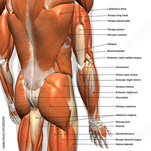
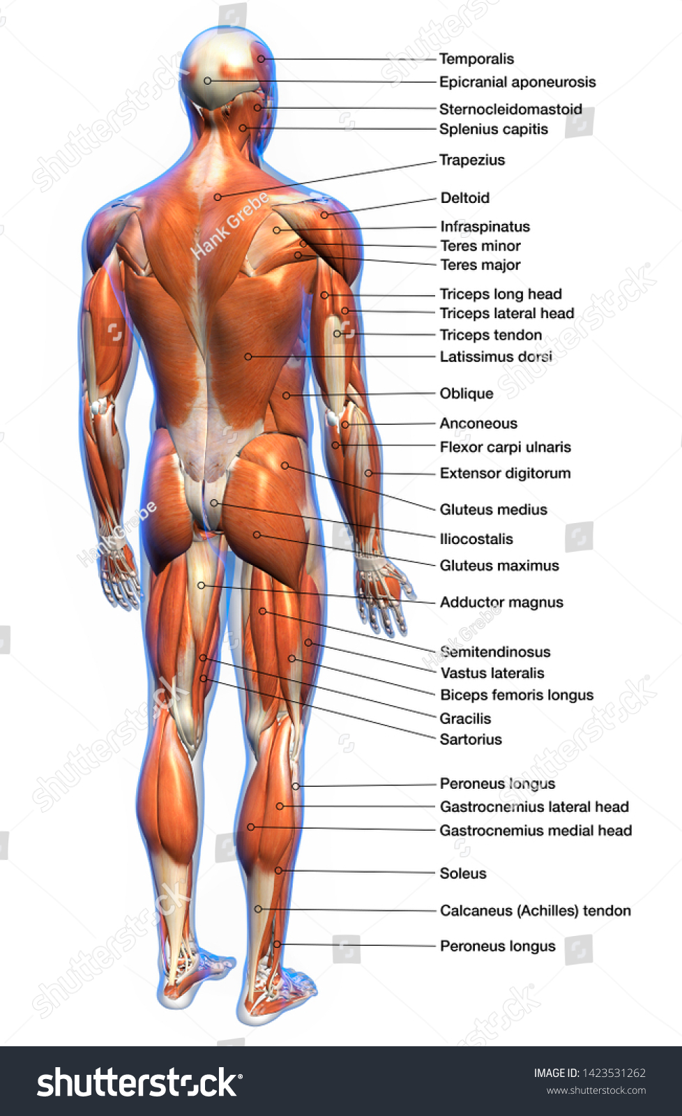
Muscular System Labeled Diagram Pictures, Images and Stock Photos Labeled human anatomy diagram of man's arm, shoulder and upper back muscles in a posterior view on a white background. Female Anterior Leg Muscles Labeled on White Front view of woman's thigh and knee muscles with names Biceps Brachii Muscles Isolated Anterior View Anatomy Labeled on...
labelled muscle diagram labelled muscle diagram 2010 Lecture 14 - Embryology. 16 Pictures about 2010 Lecture 14 - Embryology : Muscle Diagram Labeled, Muscular System - josi's Anatomy and physiology and also Printable Muscle Anatomy Chart - Muscle Anatomy Models Muscular Charts. 2010 Lecture 14 - Embryology embryology.med.unsw.edu.au
Muscle Diagram To Label Blank Muscles Diagram To Label Google Search Muscle Diagram Human Body Muscles Human Muscular System from . Label structure of skeletal muscle. Superficial and deep anterior muscles of upper body superficial and deep posterior muscles of upper body. Leversedge 2012-03-28 Pocketbook of Hand and Upper Extremity Anatomy.
Muscular System Diagram Posterior (Back) View - Sport Fitness Advisor This muscular system diagram shows the major muscle groups from the back or posterior view. To see a muscular system picture from the anterior (front) view click here. ... Muscle Anatomy & Structure. Lactic Acid, Blood Lactate & The Lactic Acid Myth. The Lactate Threshold. Hypertrophy in Human Muscle.
labeled muscles of lower leg - Pinterest Feb 16, 2015 - ... system diagram labeled 209 Human Muscular System Diagram Labeled Feb 16, 2015 - ... system diagram labeled 209 Human Muscular System Diagram Labeled Pinterest. Today. Explore. When autocomplete results are available use up and down arrows to review and enter to select. Touch device users, explore by touch or with swipe gestures.
Muscle Anatomy - Human Anatomy Chart - King of the Gym Leg, Hip & Gluteal Anatomy. Gluteal Muscles. Hamstring Muscles. Hip Adductors. Hip Flexors (Iliopsoas) Quadriceps Muscles. Neck Anatomy. Triceps Anatomy. Shoulder Anatomy (Deltoids & Rotator Cuff)
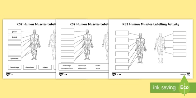
label muscles - Labelled diagram - Wordwall label muscles. Share Share by Lmorgan. KS2 Science. Show More. Like. Edit Content. Embed. More. Leaderboard. Show more Show less . This leaderboard is currently private. Click Share to make it public. This leaderboard has been disabled by the resource owner. This leaderboard is disabled as your options are different to the resource owner. ...
Labeled Muscle Diagram Chart Free Download - Formsbirds Labeled Muscle Diagram Tr a p e zius Latissimus dorsi Gluteus maximus Hamstrings Gastrocnemius Achilles P ectorals (pecs) Rectus abdominis (abs) Biiceps Ob liques Quadriceps (quads) Deltoid Tr iceps Page 1/1 Free Download Labeled Muscle Diagram Chart PDF Favor this template? Just fancy it by voting! ( 0 Votes) 0.0
Muscle Anatomy Quiz - Registered Nurse RN Muscle anatomy quiz for anatomy and physiology! When you are taking anatomy and physiology you will be required to identify major muscles in the human body. This quiz requires labeling, so it will test your knowledge on how to identify these muscles (latissimus dorsi, trapezius, deltoid, biceps brachii, triceps brachii, brachioradialis, pectoralis major, serratus anterior, rectus abdominis, etc.).
PDF Labeled Muscle Diagram - Deer Valley Unified School District Activity 4.6 Labeled Muscle Diagram From Physical Best activity guide: Middle and high school levels, 2nd edition, by NASPE, 2005, Champaign, IL: Human Kinetics. Labeled Muscle Diagram Trape zius Latissimus dorsi Gluteus maximus Hamstrings Gastrocnemius Achilles Pectorals (pecs) Rectus abdominis (abs) Biiceps Obliques Quadriceps
Muscular System Anatomy, Diagram & Function | Healthline Smooth muscle: Smooth muscle makes up the walls of hollow organs, respiratory passageways, and blood vessels. Its wavelike movements propel things through the bodily system, such as food through...
male torso muscle anatomy diagram Bodybuilding back exercises and anatomy - YouTube we have 10 Pictures about Bodybuilding back exercises and anatomy - YouTube like Collection Rigged - Male and Female Muscular System 3D Model rigged MA, muscle man | A&P.2.Skin.Bone.Muscle | Pinterest | Muscles and Medicine and also muscle upper arm color diagram - Google Search | Muscle anatomy, Body.
Muscle Charts of the Human Body — PT Direct For your reference value these charts show the major superficial and deep muscles of the human body. For your reference value these charts show the major superficial and deep muscles of the human body. ... Home › Training Design › Anatomy and Physiology › Muscle Charts of the Human Body.
Human Body Diagram - Bodytomy The torso of the human body also consists of the major muscles of our body; the pectoral muscles, the abdominal muscles, and the lateral muscle. Did You Know… ☛ While the size of the human head right from birth won't change drastically, it is the torso and the lower limbs that grow in length. Arms
shin muscles diagram muscular system muscles anatomy physiology leg nurseslabs lower muscle diagram body foot calf gastrocnemius human joint. Running Shin Splints Treated Graston Technique Sioux City chiropractor-sioux-city.com. shin leg lower splints muscles labeled anatomy muscle tibialis knee anterior tibia tendons tissue bone sioux chiropractor helena graston pain
Foot Diagram: Labeled Anatomy | Science Trends Jul 05, 2018 · The foot diagram has a complex structure made up of bones, ligaments, muscles, and tendons. Understanding the structure of the foot is best done by looking at a foot diagram where the anatomy has been labeled. If you would like to learn all the parts of the foot structure, you have come to the right place.
PDF ANATOMY OF THE MUSCULAR SYSTEM - Midland Independent School District individual muscle fibers, (b) surrounds groups of skeletal muscle fibers (fascicles), and (c) covers the muscle as a whole. 2. Name the tough connective tissue cord that serves to attach a muscle to a bone. 3. Name three types of fiber arrangements seen in skeletal muscle.







:watermark(/images/watermark_5000_10percent.png,0,0,0):watermark(/images/logo_url.png,-10,-10,0):format(jpeg)/images/overview_image/3199/IXVDf42LcYYpWEkyLwtg_hip-thigh-muscles-anterior_english.jpg)


:background_color(FFFFFF):format(jpeg)/images/library/12798/facial-muscles_english.jpg)




Post a Comment for "41 labeled muscle diagram"