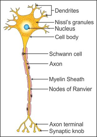
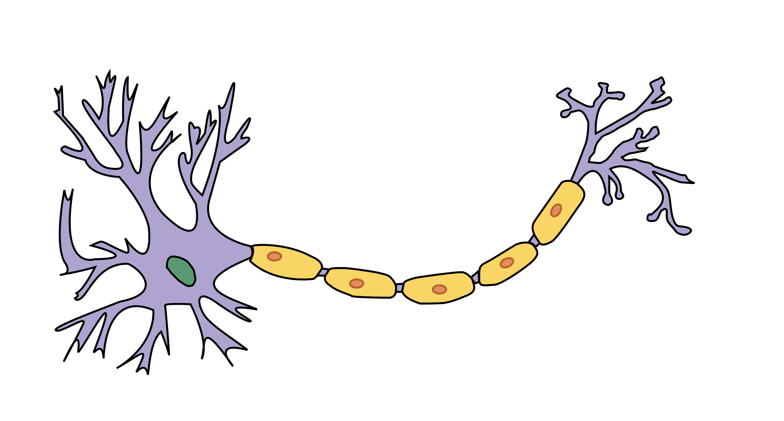
45 neuron labeling diagram
› en › productsElectronic Components and Parts Search | DigiKey Electronics Digi-Key is your authorized distributor with over a million in stock products from the world’s top suppliers. Rated #1 in content and design support! › articles › s41593/022/01041-5Single-neuron projectome of mouse prefrontal cortex | Nature ... Mar 31, 2022 · Dual-color retrograde labeling in PCG and SCm validated our single-neuron projectome results. smFISH experiment showed enriched Npnt + neurons in SCm-projecting neurons (***P = 3.5 × 10 −4 ...
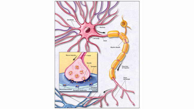
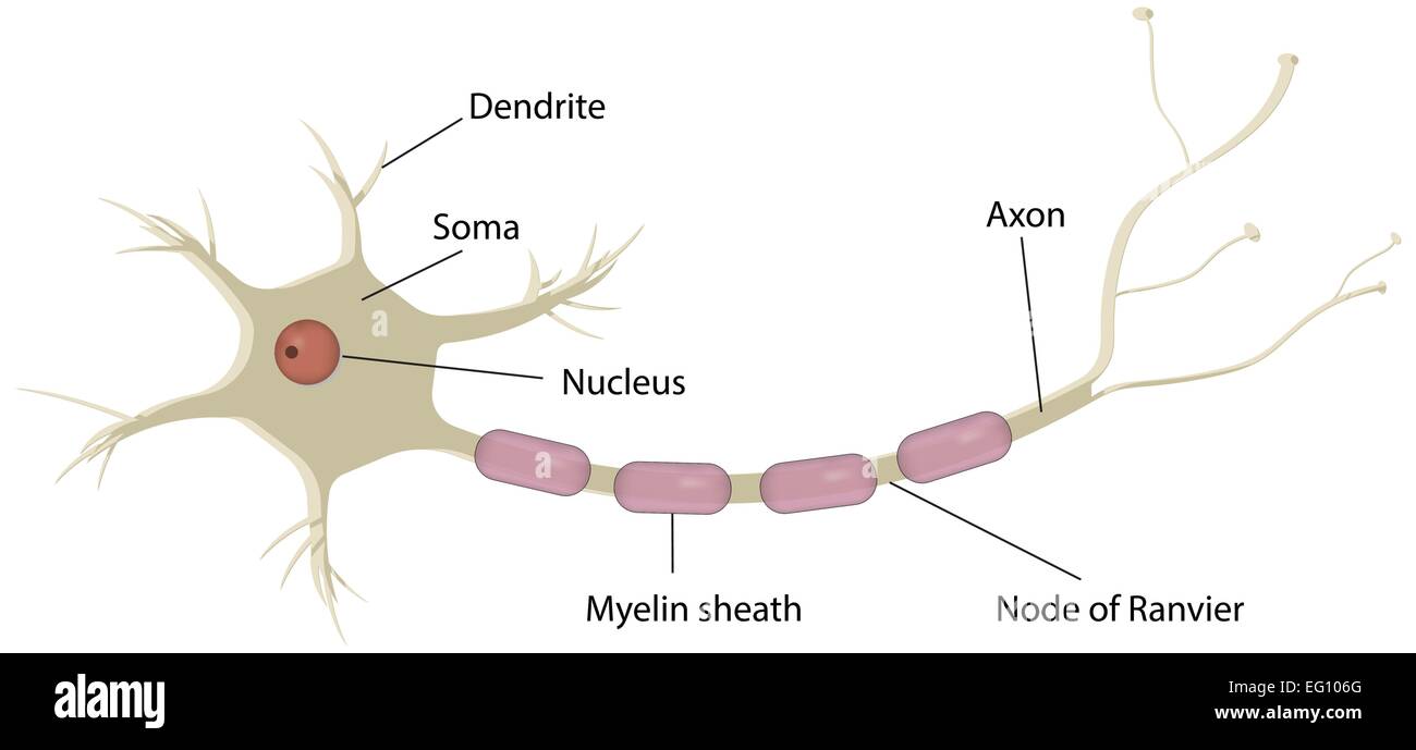
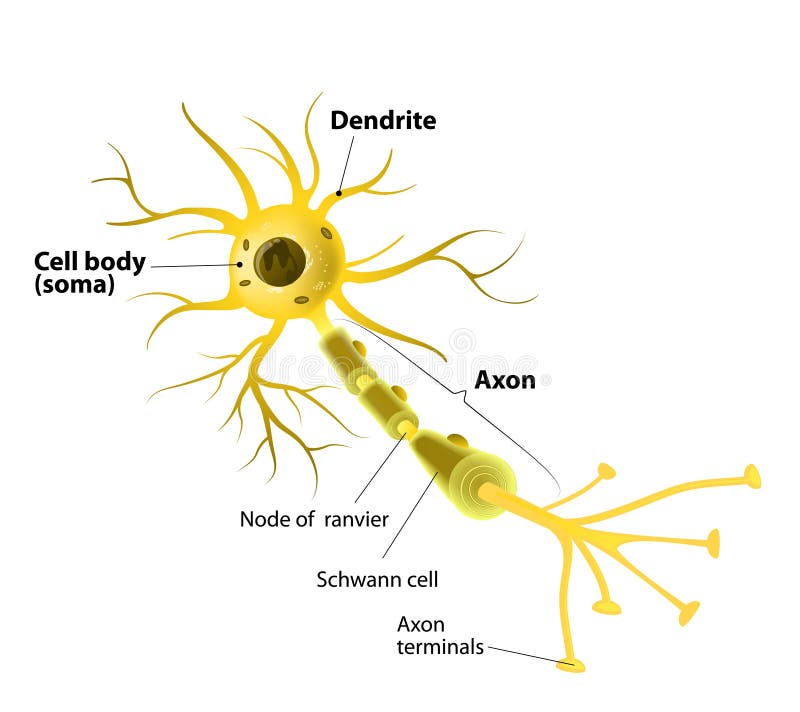
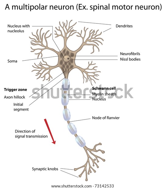
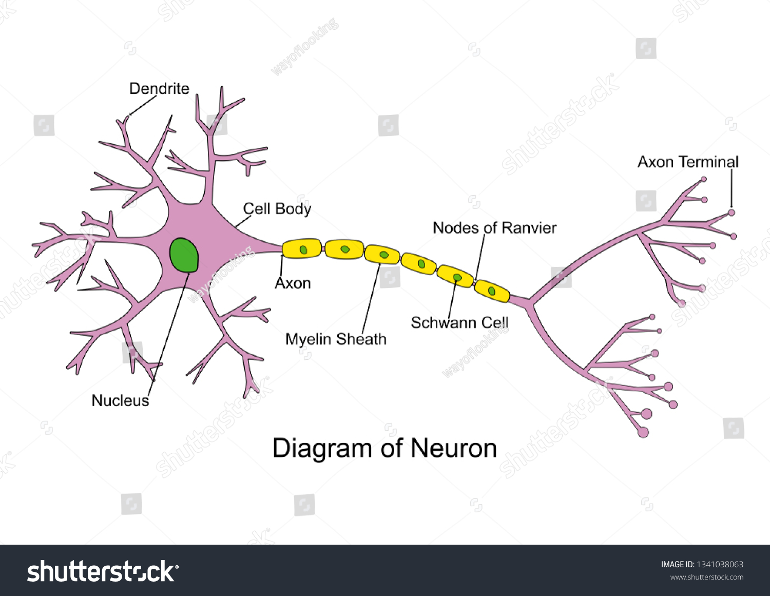
Labeled Neuron Diagram | Science Trends Labeled Neuron Diagram Alex Bolano 29, May 2019 | Last Updated: 3, March 2020 Neurons are the basic organizational units of the brain and nervous system. Neurons form the bulk of all nervous tissue and are what allow nervous tissue to conduct electrical signals that allow parts of the body to communicate with each other.
Neuron labeling diagram
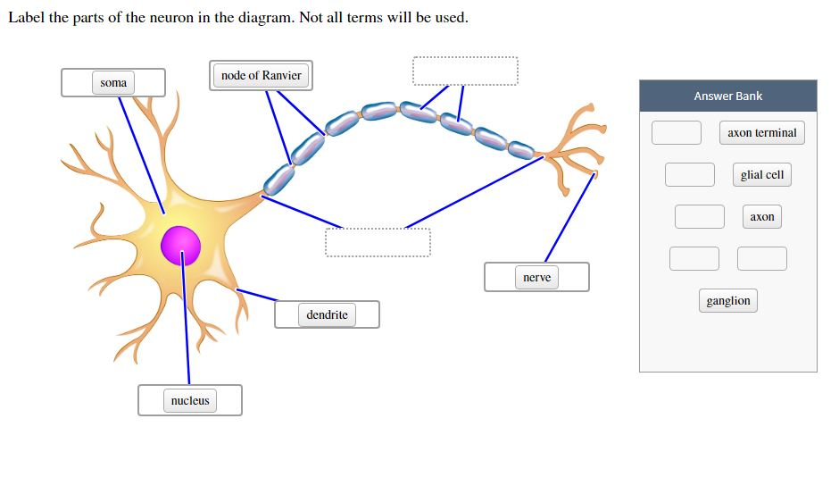
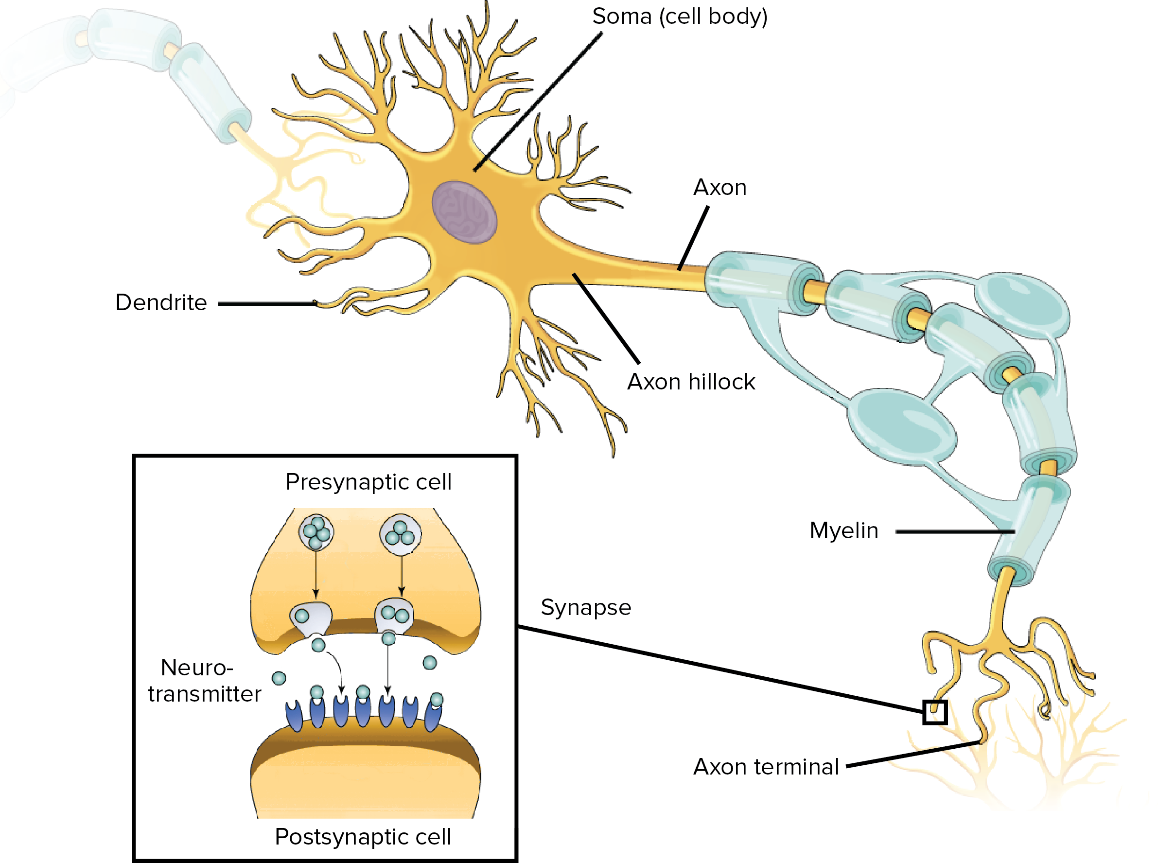
ELGi EG Series Operation And Maintenance Manual Page 53 Neuron II Manual Version : 1.8 April 2015... Page 55 INDEX Abbreviations Used 1. Technical Specification 2. Installation Instruction 2.1. Equipment’s Safety 2.1.1. Static Discharge Warning 2.1.2. Assembly 2.1.3. Do not expose to direct sunlight 2.1.4. Must be … neuron labeling Diagram | Quizlet neuron labeling Diagram | Quizlet neuron labeling + − Learn Test Match Created by leahcaroline360 PLUS Terms in this set (9) dendrite ... soma ... axon hillock ... node of Ranvier ... myelin ... telodendria ... terminal end boutons (buttons) ... glial cell ... axon ... Sets found in the same folder brain labeling 3 terms leahcaroline360 PLUS Machine Learning Glossary | Google Developers In reinforcement learning, the mechanism by which the agent transitions between states of the environment.The agent chooses the action by using a policy. activation function. A function (for example, ReLU or sigmoid) that takes in the weighted sum of all of the inputs from the previous layer and then generates and passes an output value (typically nonlinear) to the next layer.
Neuron labeling diagram. A Guide to Understand Neuron with Neuron Diagram 3.1 How to Draw a Neuron Diagram from Sketch Step 1: First, the students need to draw a circle. Based on it, they need to draw a star-like shape. It is called the cell body of the neurons. One corner of the stars is extended, forming a very thin-tube-like structure-the Axon. › manual › 1680762ELGi EG Series Operation And Maintenance Manual Page 53 Neuron II Manual Version : 1.8 April 2015... Page 55 INDEX Abbreviations Used 1. Technical Specification 2. Installation Instruction 2.1. Equipment’s Safety 2.1.1. Static Discharge Warning 2.1.2. Assembly 2.1.3. Do not expose to direct sunlight 2.1.4. Must be protected from rain 2.1.5. Peripheral nervous system: Anatomy, divisions, functions - Kenhub Aug 02, 2022 · Peripheral nerves. The workhorse of the peripheral nervous system are the peripheral nerves.Each nerve consists of a bundle of many nerve fibers and their connective tissue coverings. Each nerve fiber is an extension of a neuron whose cell body is held either within the grey matter of the CNS or within ganglia of the PNS. The comparable structure of the … Neuron Labeling Quiz - PurposeGames.com This is an online quiz called Neuron Labeling. There is a printable worksheet available for download here so you can take the quiz with pen and paper. Total. 0. Get started! Rank. --. 0. 8.
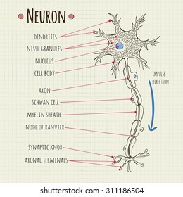
› 2015 › 06Plant Cell and Animal Cell Diagram Quiz This quiz is designed to assess your understanding of the "Difference between plant cell and animal cell".Choose the best answer from the four options given. When you've finished answering as many of the questions as you can, scroll down to the bottom of the page and check your answers by clicking 'Score'. Label Parts of a Neuron Diagram | Quizlet Dendrites. receives impulses from other nerve cells. axon hillock. The cell body...the part of the cell that houses the nucleus and keeps the entire cell alive and functioning. Myelin Sheath. Surrounds the axon an insulates it from surrounding cells and tissues and making signal transitions faster and more efficient. Terminal Buttons. Labeled Neuron Diagram| EdrawMax Template The following labeled diagram shows the parts of a neuron. In order to make it more understandable to the students, we have added all the functions of the Neuron in the labeled diagram. The major parts of the Neuron are Dendrites, Cell Body, Cell Membrane, Axon Hillock, Node of Ranvier, Schwann Cell, Axon Terminal, Myelin Sheath, Axon, and Nucleus. › books › NBK507888Neuroanatomy, Posterior Column (Dorsal Column) - StatPearls ... Jul 31, 2021 · The dorsal column, also known as the dorsal column medial lemniscus pathway, deals with the conscious appreciation of fine touch, 2-point discrimination, conscious proprioception, and vibration sensations from the body; sparing the head. In the spinal cord, this pathway travels in the dorsal column, and in the brainstem, it is transmitted through the medial lemniscus hence the name dorsal ...
Nervous system: Structure, function and diagram | Kenhub Sep 07, 2022 · Neurons, or nerve cell, are the main structural and functional units of the nervous system.Every neuron consists of a body (soma) and a number of processes (neurites). The nerve cell body contains the cellular organelles and is where neural impulses (action potentials) are generated.The processes stem from the body, they connect neurons with each other and … Plant Cell and Animal Cell Diagram Quiz - Biology Multiple … This quiz is designed to assess your understanding of the "Difference between plant cell and animal cell".Choose the best answer from the four options given. When you've finished answering as many of the questions as you can, scroll down to the bottom of the page and check your answers by clicking 'Score'. Single-neuron projectome of mouse prefrontal cortex - Nature Mar 31, 2022 · Dual-color retrograde labeling in PCG and SCm validated our single-neuron projectome results. smFISH experiment showed enriched Npnt + neurons in SCm-projecting neurons (***P = 3.5 × 10 −4 ... neuron labeling 2 Diagram | Quizlet neuron labeling 2 Diagram | Quizlet neuron labeling 2 + − Learn Test Match Created by leahcaroline360 PLUS Terms in this set (7) axon hillock ... end bouton ... post-synaptic ion channels ... neurotransmitter ... synaptic vesicles ... teleodendria ... node of Ranvier ... Sets found in the same folder brain labeling 3 terms leahcaroline360 PLUS
› food › food-labeling-nutritionChanges to the Nutrition Facts Label | FDA - U.S. Food and ... Mar 07, 2022 · There are different labeling requirements for single-ingredient sugars. The list of nutrients that are required or permitted to be declared is being updated. Vitamin D and potassium are required ...
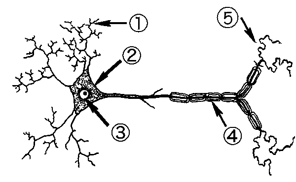
Diagram Quiz on Neuron Structure and Function (Labeling Quiz) 1. Identify the cell type in the above figure Liver Cell Cardiac Cell Nerve cell Skin cell 2. In the figure, labeled '1' receives impulses from adjacent neuron. It is called the Dendron Dendrite Axon Axonite 3. In the figure, labeled '2' is the short filaments from the cell body that carries impulses from dendrites to the cell body which is the
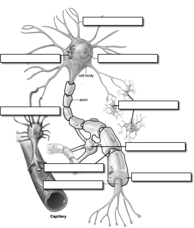
Neuron Model Labeling Diagram | Quizlet Neuron a specialized cell transmitting nerve impulses. Dendrites a short branched extension of a nerve cell, along which impulses received from other cells at synapses are transmitted to the cell body. Myelin Sheath fatty white substance that surrounds the axon of some nerve cells, forming an electrically insulating layer. Schwann Cell
Electronic Components and Parts Search | DigiKey Electronics Digi-Key is your authorized distributor with over a million in stock products from the world’s top suppliers. Rated #1 in content and design support!
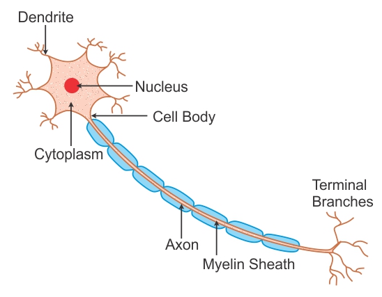
. Draw a labeled diagram of a neuron - pw.live Best Answer. Explanation: The neurons can be defined as the structures that act as the fundamental units of the nervous system. Neurons play an eminent role in receiving and sending signals to the brain. The main three parts that are present in a neuron are dendrites, cell body, and axon. The well-labeled diagram of a neuron is shown below:
Changes to the Nutrition Facts Label | FDA Mar 07, 2022 · There are different labeling requirements for single-ingredient sugars. The list of nutrients that are required or permitted to be declared is being updated. Vitamin D and potassium are required ...
Neuron Diagram & Types | Ask A Biologist - Arizona State University They pass signals from one neuron to the next inside the central nervous system. Pyramidal neurons are named after the shape of their cell body, which looks like a pyramid. They have one axon and two main dendrite branches. These cells pass signals inside the brain and tell your muscles to move.
Study Guide to the Systems of the Body - ACLS Dec 27, 2020 · Spaces are available for labeling the various parts. Skeleton match activity: Learning about the common and proper names of bones can be fun. Print this PDF and connect the common names of the bones with the proper names. ... Make a neuron out of clay by following the instructions found on this page. The directions for the model are for kids in ...
A Labelled Diagram Of Neuron with Detailed Explanations - BYJUS Diagram Of Neuron with Labels Here is the description of human neuron along with the diagram of the neuron and their parts. The neuron is a specialized and individual cell, which is also known as the nerve cell. A group of neurons forms a nerve.
Labeling A Neuron Teaching Resources | Teachers Pay Teachers You can use this resource to label parts of a neuron as a model, diagram, or notes in your nervous system unit to instruct, explain, and facilitate student learning about neurons' design in the human body. ... Color Diagrams, Labeling and Coloring Activities for high school and college anatomy students, including: reading, color diagrams ...
diagram of a neuron labeled Neuron labeled neurone labelled disease guillain hirn sklerose nervensystem schmerzen naturheilkunde Nervous tissue. 17 Images about Nervous tissue : Neuron Labeled Diagram Stock Vector Art & Illustration, Vector Image, Conditioning, Patterns and Happiness - Maximising Your Happiness Part 6 and also Events at the Neuromuscular Junction - YouTube.
Neurons (With Diagram) - Biology Discussion A neuron is a structural and functional unit of the neural tissue and hence the neural system. Certain neurons may almost equal the length of body itself. Thus neurons with longer processes (projections) are the longest cells in the body. Human neural system has about 100 billion neurons. Majority of the neurons occur in the brain.
Neuron under Microscope with Labeled Diagram - AnatomyLearner Let's see the neuron histology slide labelled diagram and try to find out the below-mentioned characteristics - Presence of an identifiable cell body (soma) that locates in the brain's grey matter (according to the slide image). The cell body possesses spherical, euchromatic, and large eccentric nuclei containing a prominent nucleolus.
General Structure of a Neuron (Nerve Cell) | GetBodySmart General Structure of a Neuron (Nerve Cell) Start Quiz. Learn this topic from scratch or practice what you already know with these interactive spaced repetition-inspired anatomy quizzes. Learn anatomy faster and. remember everything you learn. Start Now. <.
› en › libraryNervous system: Structure, function and diagram | Kenhub Sep 07, 2022 · Nervous system breakdown (diagram) So nervous tissue, comprised of neurons and neuroglia, forms our nervous organs (e.g. the brain, nerves). These organs unite according to their common function, forming the evolutionary perfection that is our nervous system. The nervous system (NS) is structurally broken down into two divisions;
Label Neuron Anatomy Printout - EnchantedLearning.com Read the definitions, then label the neuron diagram below. axon - the long extension of a neuron that carries nerve impulses away from the body of the cell. cell body - the cell body of the neuron; it contains the nucleus (also called the soma) dendrites - the branching structure of a neuron that receives messages (attached to the cell body)
Neuroanatomy, Posterior Column (Dorsal Column) - NCBI Bookshelf Jul 31, 2021 · The dorsal column, also known as the dorsal column medial lemniscus pathway, deals with the conscious appreciation of fine touch, 2-point discrimination, conscious proprioception, and vibration sensations from the body; sparing the head. In the spinal cord, this pathway travels in the dorsal column, and in the brainstem, it is transmitted through the medial …
Neuron Diagram Labeled | EdrawMax Template It is an effective form of self-assessment, enabling students to check their understanding. In the following diagram, we have illustrated the important parts of the Neuron. In the following Neuron labeled diagram, we have dendrite, cell body, axon, myelin sheath, Schwann cell, a node of Ranvier, axon terminal, and nucleus.
labeling a neuron Diagram | Quizlet labeling a neuron Diagram | Quizlet labeling a neuron + − Learn Test Match Created by elliezap PLUS Terms in this set (10) node of Ranvier ... dendrite ... telodendria ... neurotransmitters ... synaptic vesicles ... terminal end button ... axon hillock ... myelin sheath ... soma ... receptor site ... elliezap elliezap
Describe the structure and function of neuron with labelled diagram ... 6.At the terminals, depending on the makeup of the neuron, the impulse is transmitted to the dendrites of the next neuron via a junction known as synapse, in a chemical or electric manner Diagram of neuron with labels. Here is the description of human neuron along with the diagram of the neuron and their parts.
Machine Learning Glossary | Google Developers In reinforcement learning, the mechanism by which the agent transitions between states of the environment.The agent chooses the action by using a policy. activation function. A function (for example, ReLU or sigmoid) that takes in the weighted sum of all of the inputs from the previous layer and then generates and passes an output value (typically nonlinear) to the next layer.
neuron labeling Diagram | Quizlet neuron labeling Diagram | Quizlet neuron labeling + − Learn Test Match Created by leahcaroline360 PLUS Terms in this set (9) dendrite ... soma ... axon hillock ... node of Ranvier ... myelin ... telodendria ... terminal end boutons (buttons) ... glial cell ... axon ... Sets found in the same folder brain labeling 3 terms leahcaroline360 PLUS
ELGi EG Series Operation And Maintenance Manual Page 53 Neuron II Manual Version : 1.8 April 2015... Page 55 INDEX Abbreviations Used 1. Technical Specification 2. Installation Instruction 2.1. Equipment’s Safety 2.1.1. Static Discharge Warning 2.1.2. Assembly 2.1.3. Do not expose to direct sunlight 2.1.4. Must be …

















Post a Comment for "45 neuron labeling diagram"