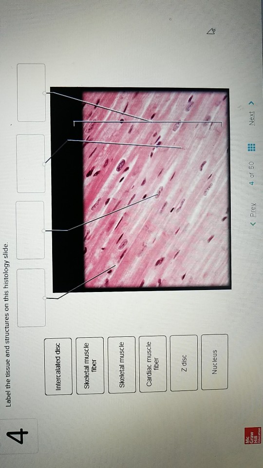
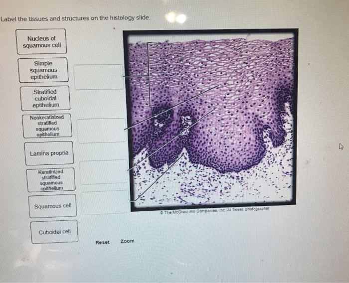
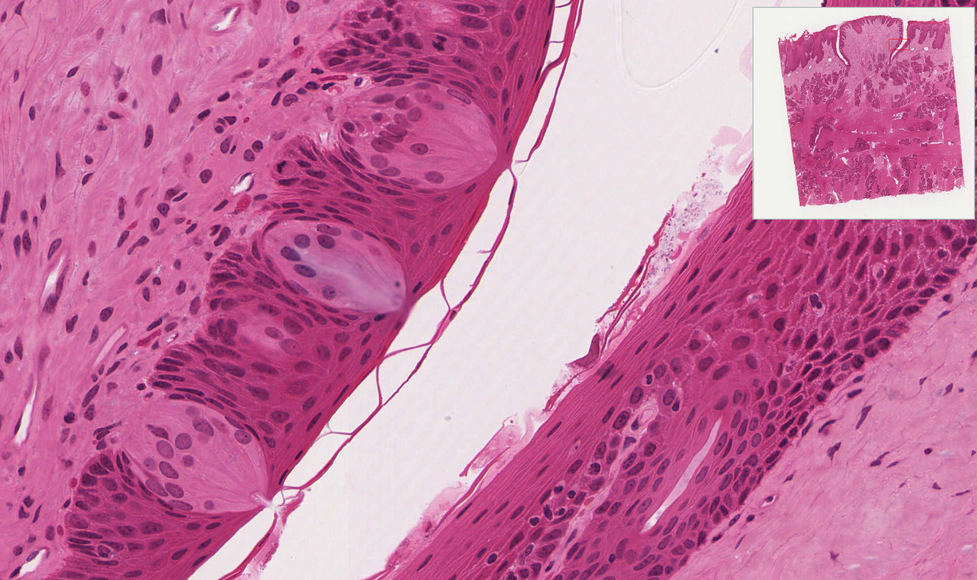
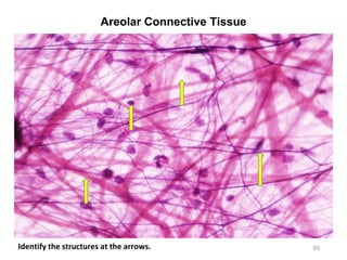
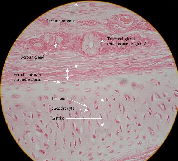
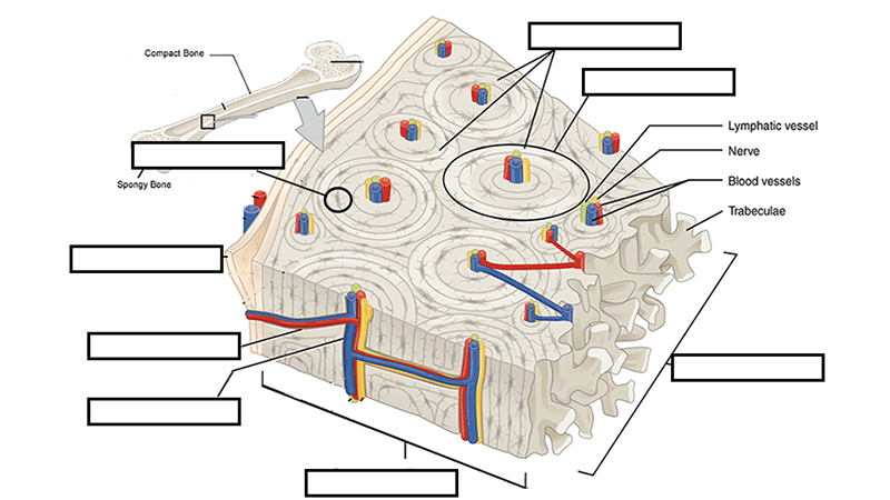
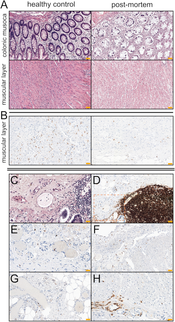
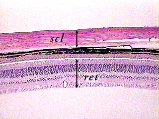
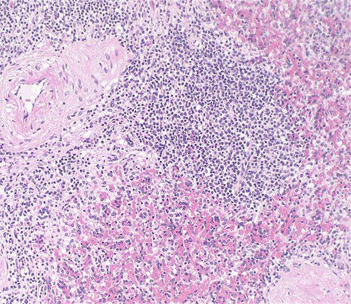
44 label the tissue and structures on this histology slide
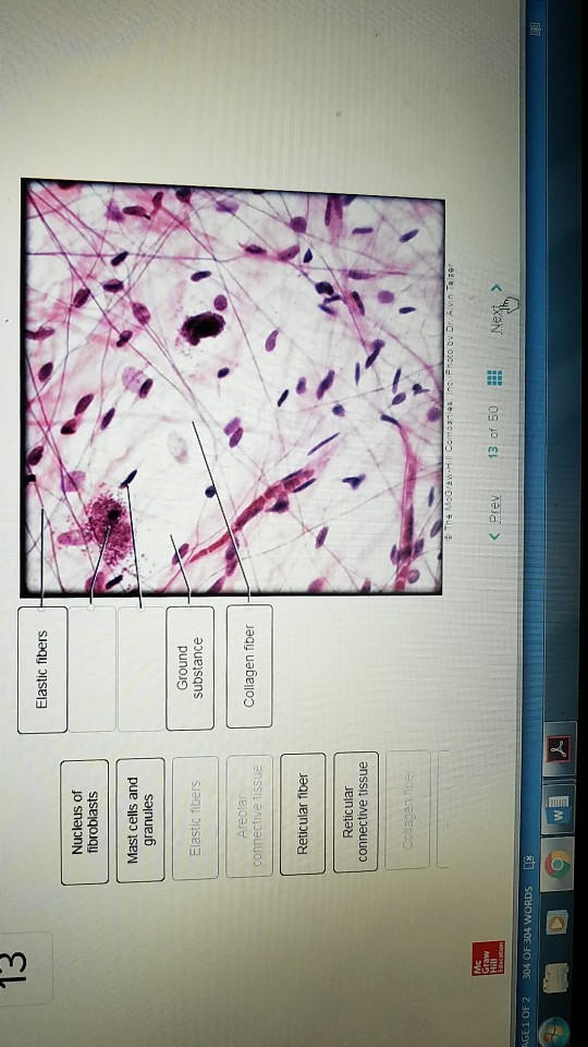
answer asap pleaseeee Label the tissues and structures on the... Label the tissue and structures on this histology slide. Collagen fiber Areolar connective tissue Elastic fibers Mast cells and granules Nucleus of fibroblasts Reticular fiber Ground substance Reticular @ The McGraw-Hill Companies, Inc./Photo by Dr. Alvin Telser connective tissue Name the tissue tune found on this slide: Artery Histology - Elastic and Muscular Arteries Slides - AnatomyLearner Okay, let's identify the muscular artery histology slide under a light microscope with the following identifying characteristics. The sample tissue section shows the three different layers - tunica intima, tunica media, and tunica externa or adventitia. The tunica intima is lined by the simple squamous epithelium (endothelium).
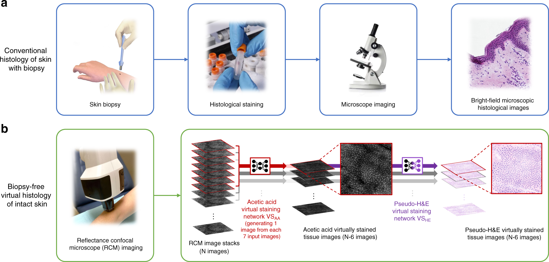
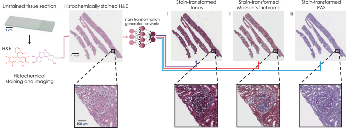
Histology: Stains and section interpretation | Kenhub A huge range of stains is used in histology, from dyes and metals to labeled antibodies. Certain stains change the coloration of cells and tissues significantly, different from the color of the original dye complex, a phenomenon known as metachromasia. For staining, paraffin sections of tissue are normally used.
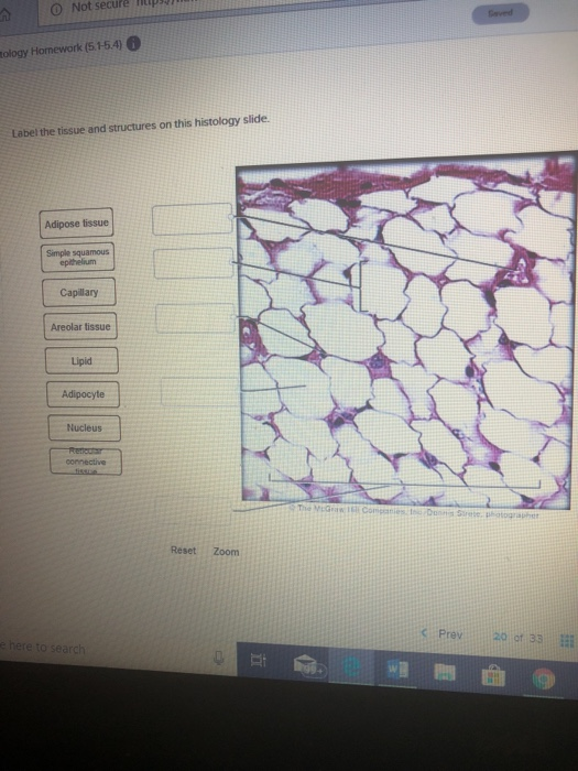
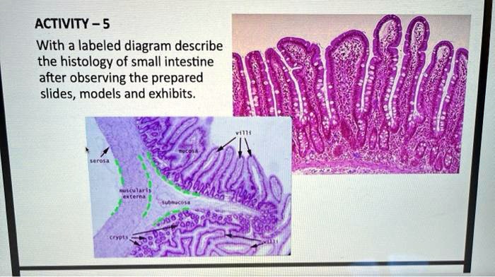
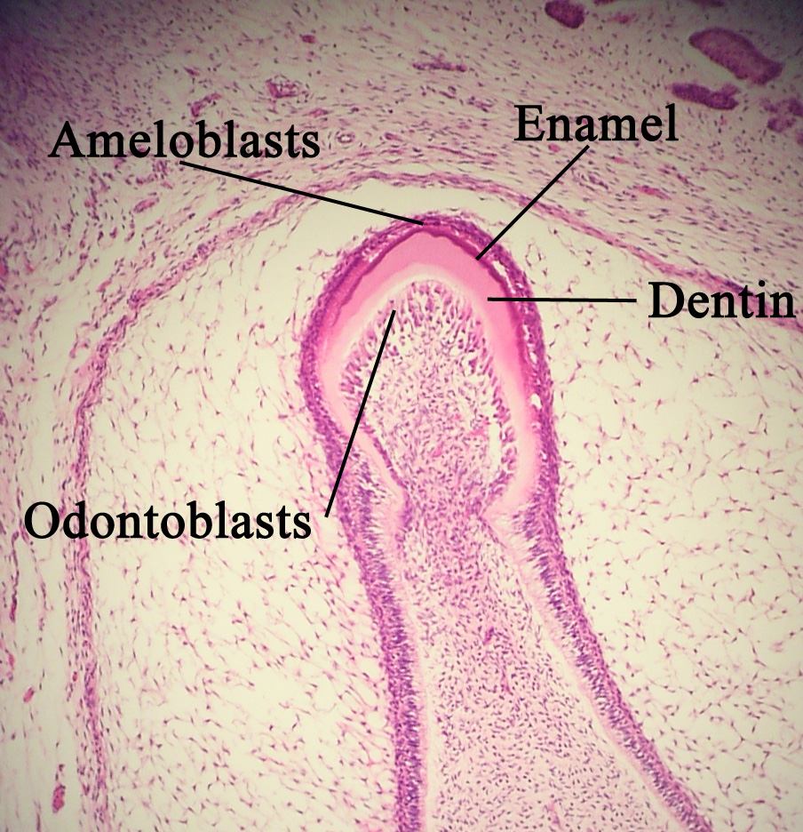
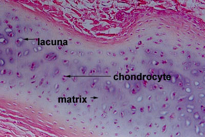
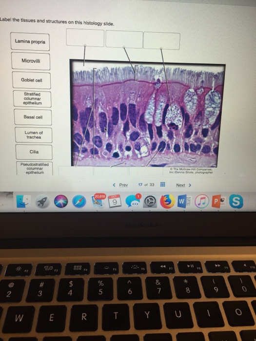
Label the tissue and structures on this histology slide
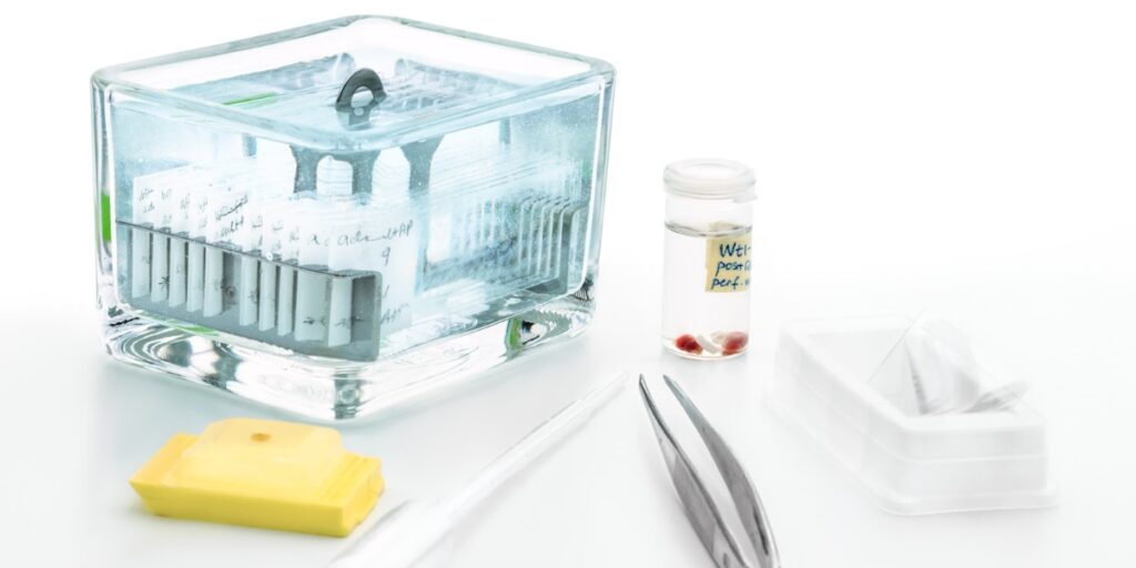
Histology Slide Preparation: 5 Simple Steps - Bitesize Bio The Five Steps of Histology Slide Preparation. 1. Tissue fixation. Slide preparation begins with the fixation of your tissue specimen. This is a crucial step in tissue preparation, and its purpose is to prevent tissue autolysis and putrefaction. For best results, your biological tissue samples should be transferred into fixative immediately ... How to examine histology slides: Techniques and tips | Kenhub How to examine histology slides Author: Alexandra Sieroslawska MD • Reviewer: Dimitrios Mytilinaios MD, PhD Last reviewed: September 14, 2022 Reading time: 3 minutes Histology is a beautiful subject that allows us to explore the structure and function of tissues. If we take a little sample of an organ tissue, stain it accordingly and place it under a light microscope, we are able to see the ... Microscope Slides of Cells and Tissues | Histology Guide Organs are assembled from the four basic types of tissues and have cells with specialized functions. Chapter 9. Cardiovascular System. Chapter 10. Lymphoid System. Chapter 11. Skin. Chapter 12. Exocrine Glands.
Label the tissue and structures on this histology slide. Need help identifying tissues? Try our tissue quizzes! | Kenhub For example, our connective tissue quizzes with pictures show you a series of histology slides and challenge you to identify the correct structure out of up to 4 images, based on text prompts. If you think you've got the hang of it pretty well, challenge yourself with the advanced tissue identification quizzes, which don't contain prompts. Skeletal Muscle Histology Slide Identification and Labeled Diagram ... Don't forget to read the other articles from anatomy learner (related to animal anatomy or histology) - #1. Details guide and identification points of smooth muscle histology #2. Dense connective tissue identification points with real slide images #3. Loose connective tissue slide identification. Conclusion Solved Label the tissue and structures on this histology - Chegg Expert Answer. The reticular connective tissue consists of network of the fine reticul …. View the full answer. Transcribed image text: Label the tissue and structures on this histology slide. Areolar connective 0 tissue Areolar connective tissue Leukocyte Reticular connective tissue Reticular connective tissue Leukocyte Ground substance ... Solved Label the tissues and structures on this histology - Chegg This problem has been solved! You'll get a detailed solution from a subject matter expert that helps you learn core concepts. Question: Label the tissues and structures on this histology slide. Lamina propria Goblet cell Stratified columnar epithelium Basal cell Lumen of Cilia The Mcüraw < Prev 170' 33Ⅲ Next > 名0 2 3 4.
Histology Lab Photo Quiz Flashcards | Quizlet Sets found in the same folder. BIOL 121 Appendicular Bones and Bony Landmarks. 173 terms Images. robswatski Teacher. Histology Lab Photo Quiz. 21 terms Images. csaluki762. BIOL 121 Axial Skeleton Bones and Bony Landma…. 220 terms Images. Histology Slides Identification from Different Organ Systems Muscular tissue histology slide. Three types of muscle histology slides can be identified with their special morphology or structure under the light microscope. All muscle tissue contains the elongated cell (known as fibers). Here, you should learn and identify the three types of muscle tissue histology slides with identification points. #1. Tissue slides Flashcards | Quizlet Elastic fibers (thin) Adipose (loose) CT. Addi poses on ROCKS. Similar to areolar in structure, but w/ ADIPOCYTES (fat cells) filled w/ oil droplet. This tissue wraps & cushions organs, insulates & energy storage. Location: Under skin in subcutaneous tissue, around kidneys & eyeballs, w/in abdomen, in breasts. Label: Histology guide: Definition and slides | Kenhub Human anatomy is pretty straightforward. If you were to look at some bones on a skeleton, you'd see a greyish rigid mass with some bumps and depressions. However, if you take a much closer look, you'll see that the histology of bones, is a whole other story. Histology is the science of the microscopic structure of cells, tissues and organs.
histology human anatomy tissue slides - Quizlet epithelial tissue histology slides. stratified squamous epithelium. simple ciliated columnar epithelium. pseudostratified ciliated columnar epit…. flattened tile-like cells in surface layer, rounder cells in b…. elongated cells, oval shaped nuclei, single layer, projections…. Actually a single layer of cells of varying height, some not r ... tissue histology slides Flashcards | Quizlet tissue histology slides. 4.8 (13 reviews) Flashcards. Learn. ... BIO 150- Human Anatomy Lab 2 Skeletal System. 61 terms. Images. toriimamie. Tissue Histology Slides. 35 terms. Images. elisacarter. A&P Lab Histology Slides. 20 terms. Images. Lachin_gilman. Recent flashcard sets. Antimicrobial drugs part 1. Solved Label the tissues and structures on the histology - Chegg Expert Answer. 100% (4 ratings) Squamous cell : Thin, flat cells found in tissues that form surface of skin, lining of hollow organs of body and lining of respiratory and di …. View the full answer. Transcribed image text: Label the tissues and structures on the histology slide. Squamous cell Nucleus of squamous cell Keratinized stratified ... Histology Slides Flashcards | Quizlet Study with Quizlet and memorize flashcards containing terms like Tissue: Aerolar Location: Surrounds capillaries Function: wraps and cushions organs Structures: Collagen fibers, elastine fibers, fibroblast nuclei Things to pay attention to: Hints:, Tissue:Adipose Location: under skin, breasts Function: insulates against heat loss Structures: nucleus, fat storage area Things to pay attention to ...
Microscope Slides of Cells and Tissues | Histology Guide Organs are assembled from the four basic types of tissues and have cells with specialized functions. Chapter 9. Cardiovascular System. Chapter 10. Lymphoid System. Chapter 11. Skin. Chapter 12. Exocrine Glands.
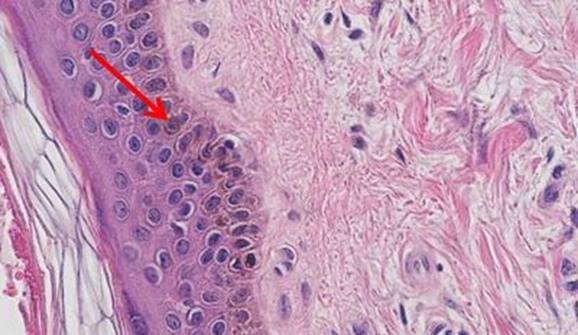
How to examine histology slides: Techniques and tips | Kenhub How to examine histology slides Author: Alexandra Sieroslawska MD • Reviewer: Dimitrios Mytilinaios MD, PhD Last reviewed: September 14, 2022 Reading time: 3 minutes Histology is a beautiful subject that allows us to explore the structure and function of tissues. If we take a little sample of an organ tissue, stain it accordingly and place it under a light microscope, we are able to see the ...
Histology Slide Preparation: 5 Simple Steps - Bitesize Bio The Five Steps of Histology Slide Preparation. 1. Tissue fixation. Slide preparation begins with the fixation of your tissue specimen. This is a crucial step in tissue preparation, and its purpose is to prevent tissue autolysis and putrefaction. For best results, your biological tissue samples should be transferred into fixative immediately ...




:watermark(/images/watermark_only_sm.png,0,0,0):watermark(/images/logo_url_sm.png,-10,-10,0):format(jpeg)/images/anatomy_term/gastric-body-3/ZmAOsYiaMqAumIIHVP9eg_Gastric_Body.png)









Post a Comment for "44 label the tissue and structures on this histology slide"